 Movie
Movie Controller

[English] 日本語
 Yorodumi
- PDB-3t5i: Structure of Fully modified farnesylated Rheb Peptide in complex ... -
+ Open data

- Basic information
| Entry | Database: PDB / ID: 3t5i | ||||||
|---|---|---|---|---|---|---|---|
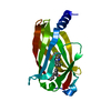
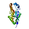
| Title | Structure of Fully modified farnesylated Rheb Peptide in complex with PDE6D | ||||||
 Components Components |
| ||||||
 Keywords Keywords | LIPID BINDING PROTEIN / Immunoglobulin-like beta sandwitch fold / Rheb / Farnesyl / prenyl / SIGNALING PROTEIN | ||||||
| Function / homology |  Function and homology information Function and homology informationARL13B-mediated ciliary trafficking of INPP5E / GTPase inhibitor activity / visual perception / cytoplasmic vesicle membrane / small GTPase binding / RAS processing / cytoplasmic vesicle / cilium / cytosol / cytoplasm Similarity search - Function | ||||||
| Biological species |  Homo sapiens (human) Homo sapiens (human) | ||||||
| Method |  X-RAY DIFFRACTION / SYNCHROTRON / MOLECULAR REPLACEMENT / Resolution: 2.1 Å X-RAY DIFFRACTION / SYNCHROTRON / MOLECULAR REPLACEMENT / Resolution: 2.1 Å | ||||||
 Authors Authors | Ismail, S.A. / Chen, Y.-X. / Wittinghofer, A. | ||||||
 Citation Citation | Journal: Nat.Chem.Biol. / Year: 2011 Title: Arl2-GTP and Arl3-GTP regulate a GDI-like transport system for farnesylated cargo. Authors: Ismail, S.A. / Chen, Y.X. / Rusinova, A. / Chandra, A. / Bierbaum, M. / Gremer, L. / Triola, G. / Waldmann, H. / Bastiaens, P.I. / Wittinghofer, A. | ||||||
| History |
|
- Structure visualization
| Structure viewer | Molecule: MolmilJmol/JSmol |
|---|
- Downloads & links
-Download
| PDBx/mmCIF format | 3t5i.cif.gz | 138.6 KB | Display | PDBx/mmCIF format |
|---|---|---|---|---|
| PDB format | pdb3t5i.ent.gz | 109.4 KB | Display | PDB format |
| PDBx/mmJSON format | 3t5i.json.gz | Tree view | PDBx/mmJSON format | |
| Others |  Other downloads Other downloads |
-Validation report
| Arichive directory | https://data.pdbj.org/pub/pdb/validation_reports/t5/3t5iftp://data.pdbj.org/pub/pdb/validation_reports/t5/3t5i | HTTPS FTP |
|---|
-Related structure data
| Related structure data |  3t5gSC S: Starting model for refinement C: citing same article ( |
|---|---|
| Similar structure data |


-Links
 PDBj
PDBj- Assembly
| Deposited unit | 
| ||||||||
|---|---|---|---|---|---|---|---|---|---|
| 1 | 
| ||||||||
| 2 | 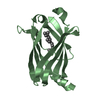
| ||||||||
| 3 | 
| ||||||||
| 4 | 
| ||||||||
| Unit cell |
|
-Components
| #1: Protein | Mass: 17585.121 Da / Num. of mol.: 4 / Fragment: Full length PDE delta Source method: isolated from a genetically manipulated source Source: (gene. exp.) Homo sapiens (human) / Gene: PDE6D, PDED / Production host:  #2: Protein/peptide | Mass: 942.051 Da / Num. of mol.: 2 / Fragment: C-terminal Farnesylated Rheb peptide / Source method: obtained synthetically Details: Peptide synthesis , the sequence is the Cterminal sequence of Rheb #3: Chemical | ChemComp-FAR /   Mass: 206.367 Da / Num. of mol.: 4 / Source method: obtained synthetically / Formula: C15H26 Mass: 206.367 Da / Num. of mol.: 4 / Source method: obtained synthetically / Formula: C15H26#4: Water | ChemComp-HOH / |  Mass: 18.015 Da / Num. of mol.: 278 / Source method: isolated from a natural source / Formula: H2O Mass: 18.015 Da / Num. of mol.: 278 / Source method: isolated from a natural source / Formula: H2O |
|---|
-Experimental details
-Experiment
| Experiment | Method: X-RAY DIFFRACTION / Number of used crystals: 1 |
|---|
- Sample preparation
| Crystal | Density Matthews: 2.25 Å3/Da / Density % sol: 45.25 % |
|---|---|
| Crystal grow | Temperature: 293 K / Method: vapor diffusion, hanging drop / pH: 7.2 Details: 0.1 MES 6.5 and 30 % PEG 5000 MME , pH 7.2, VAPOR DIFFUSION, HANGING DROP, temperature 293K |
-Data collection
| Diffraction | Mean temperature: 100 K |
|---|---|
| Diffraction source | Source: SYNCHROTRON / Site: SLS  / Beamline: X10SA / Wavelength: 0.979 Å / Beamline: X10SA / Wavelength: 0.979 Å |
| Detector | Type: MARMOSAIC 225 mm CCD / Detector: CCD / Date: Jul 31, 2010 |
| Radiation | Monochromator: SAGITALLY - HORIZONTALLY FOCUSED SI(111) MONOCHROMATOR Protocol: SINGLE WAVELENGTH / Monochromatic (M) / Laue (L): M / Scattering type: x-ray |
| Radiation wavelength | Wavelength: 0.979 Å / Relative weight: 1 |
| Reflection | Resolution: 2.1→30 Å / Num. obs: 37401 / % possible obs: 99.6 % / Observed criterion σ(F): 1 / Observed criterion σ(I): 1 |
| Reflection shell | Resolution: 2.1→2.2 Å / % possible all: 99.5 |
- Processing
| Software |
| |||||||||||||||||||||||||||||||||||||||||||||||||||||||||||||||||
|---|---|---|---|---|---|---|---|---|---|---|---|---|---|---|---|---|---|---|---|---|---|---|---|---|---|---|---|---|---|---|---|---|---|---|---|---|---|---|---|---|---|---|---|---|---|---|---|---|---|---|---|---|---|---|---|---|---|---|---|---|---|---|---|---|---|---|
| Refinement | Method to determine structure: MOLECULAR REPLACEMENT Starting model: 3T5G Resolution: 2.1→29.91 Å / Cor.coef. Fo:Fc: 0.951 / Cor.coef. Fo:Fc free: 0.916 / SU B: 4.965 / SU ML: 0.135 / Cross valid method: THROUGHOUT / σ(F): 1 / ESU R Free: 0.199 / Stereochemistry target values: MAXIMUM LIKELIHOOD / Details: HYDROGENS HAVE BEEN ADDED IN THE RIDING POSITIONS
| |||||||||||||||||||||||||||||||||||||||||||||||||||||||||||||||||
| Solvent computation | Ion probe radii: 0.8 Å / Shrinkage radii: 0.8 Å / VDW probe radii: 1.4 Å / Solvent model: MASK | |||||||||||||||||||||||||||||||||||||||||||||||||||||||||||||||||
| Displacement parameters | Biso mean: 28.721 Å2
| |||||||||||||||||||||||||||||||||||||||||||||||||||||||||||||||||
| Refinement step | Cycle: LAST / Resolution: 2.1→29.91 Å
| |||||||||||||||||||||||||||||||||||||||||||||||||||||||||||||||||
| Refine LS restraints |
| |||||||||||||||||||||||||||||||||||||||||||||||||||||||||||||||||
| LS refinement shell | Resolution: 2.1→2.154 Å / Total num. of bins used: 20
|
