 Movie
Movie Controller


+ Open data

- Basic information
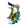
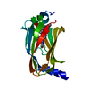
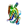
| Entry | Database: PDB / ID: 5x72 | ||||||
|---|---|---|---|---|---|---|---|
| Title | The crystal Structure PDE delta in complex with (rac)-p9 | ||||||
 Components Components | Retinal rod rhodopsin-sensitive cGMP 3',5'-cyclic phosphodiesterase subunit delta | ||||||
 Keywords Keywords | LIPID BINDING PROTEIN | ||||||
| Function / homology |  Function and homology information Function and homology informationARL13B-mediated ciliary trafficking of INPP5E / GTPase inhibitor activity / visual perception / cytoplasmic vesicle membrane / small GTPase binding / RAS processing / cytoplasmic vesicle / cilium / cytoplasm / cytosol Similarity search - Function | ||||||
| Biological species |  Homo sapiens (human) Homo sapiens (human) | ||||||
| Method |  X-RAY DIFFRACTION / SYNCHROTRON / MOLECULAR REPLACEMENT / Resolution: 1.95 Å X-RAY DIFFRACTION / SYNCHROTRON / MOLECULAR REPLACEMENT / Resolution: 1.95 Å | ||||||
 Authors Authors | Jiang, Y. / Zhuang, C. / Chen, L. / Wang, R. / Wang, F. / Sheng, C. | ||||||
| Funding support |  China, 1items China, 1items
| ||||||
 Citation Citation | Journal: J. Med. Chem. / Year: 2017 Title: Structural Biology-Inspired Discovery of Novel KRAS-PDE delta Inhibitors Authors: Jiang, Y. / Zhuang, C. / Chen, L. / Lu, J. / Dong, G. / Miao, Z. / Zhang, W. / Li, J. / Sheng, C. | ||||||
| History |
|
- Structure visualization
| Structure viewer | Molecule: MolmilJmol/JSmol |
|---|
- Downloads & links
-Download
| PDBx/mmCIF format | 5x72.cif.gz | 80.4 KB | Display | PDBx/mmCIF format |
|---|---|---|---|---|
| PDB format | pdb5x72.ent.gz | 59.5 KB | Display | PDB format |
| PDBx/mmJSON format | 5x72.json.gz | Tree view | PDBx/mmJSON format | |
| Others |  Other downloads Other downloads |
-Validation report
| Arichive directory | https://data.pdbj.org/pub/pdb/validation_reports/x7/5x72ftp://data.pdbj.org/pub/pdb/validation_reports/x7/5x72 | HTTPS FTP |
|---|
-Related structure data
| Related structure data |  5x73C  5x74C  4jv6S C: citing same article ( S: Starting model for refinement |
|---|---|
| Similar structure data |


-Links
 PDBj
PDBj- Assembly
| Deposited unit | 
| ||||||||
|---|---|---|---|---|---|---|---|---|---|
| 1 |
| ||||||||
| Unit cell |
| ||||||||
| Components on special symmetry positions |
|
-Components
| #1: Protein | Mass: 17440.990 Da / Num. of mol.: 1 Source method: isolated from a genetically manipulated source Source: (gene. exp.) Homo sapiens (human) / Gene: PDE6D, PDED / Plasmid: pET28a / Production host:  |
|---|---|
| #2: Chemical | ChemComp-P59 / (  Mass: 318.344 Da / Num. of mol.: 1 / Source method: obtained synthetically / Formula: C20H15FN2O Mass: 318.344 Da / Num. of mol.: 1 / Source method: obtained synthetically / Formula: C20H15FN2O |
| #3: Chemical | ChemComp-P69 / (  Mass: 318.344 Da / Num. of mol.: 1 / Source method: obtained synthetically / Formula: C20H15FN2O Mass: 318.344 Da / Num. of mol.: 1 / Source method: obtained synthetically / Formula: C20H15FN2O |
| #4: Water | ChemComp-HOH /  Mass: 18.015 Da / Num. of mol.: 86 / Source method: isolated from a natural source / Formula: H2O Mass: 18.015 Da / Num. of mol.: 86 / Source method: isolated from a natural source / Formula: H2O |
-Experimental details
-Experiment
| Experiment | Method: X-RAY DIFFRACTION / Number of used crystals: 1 |
|---|
- Sample preparation
| Crystal | Density Matthews: 2.95 Å3/Da / Density % sol: 58.31 % |
|---|---|
| Crystal grow | Temperature: 277 K / Method: vapor diffusion, sitting drop Details: 0.2 M Ammonium citrate dibasic, 20% w/v Polyethylene glycol 3350 |
-Data collection
| Diffraction | Mean temperature: 100 K |
|---|---|
| Diffraction source | Source: SYNCHROTRON / Site: SSRF / Beamline: BL17U / Wavelength: 0.97915 Å |
| Detector | Type: ADSC QUANTUM 315r / Detector: AREA DETECTOR / Date: Dec 18, 2015 |
| Radiation | Protocol: SINGLE WAVELENGTH / Monochromatic (M) / Laue (L): M / Scattering type: x-ray |
| Radiation wavelength | Wavelength: 0.97915 Å / Relative weight: 1 |
| Reflection | Resolution: 1.95→50 Å / Num. obs: 15663 / % possible obs: 99.7 % / Redundancy: 13.9 % / Rmerge(I) obs: 0.066 / Rpim(I) all: 0.019 / Χ2: 0.975 / Net I/σ(I): 38.61 |
| Reflection shell | Resolution: 1.95→1.98 Å / Redundancy: 14.2 % / Rmerge(I) obs: 0.481 / Mean I/σ(I) obs: 7.4 / Num. unique obs: 774 / Rpim(I) all: 0.132 / Χ2: 0.935 / % possible all: 100 |
- Processing
| Software |
| ||||||||||||||||||||||||||||||||||||||||||||||||||||||||||||||||||||||||||||||||||||||||||||||||||||||||||||||||||||||||||||||||||||||||||||||||||||||||||||||||||||||||||||||||||||||
|---|---|---|---|---|---|---|---|---|---|---|---|---|---|---|---|---|---|---|---|---|---|---|---|---|---|---|---|---|---|---|---|---|---|---|---|---|---|---|---|---|---|---|---|---|---|---|---|---|---|---|---|---|---|---|---|---|---|---|---|---|---|---|---|---|---|---|---|---|---|---|---|---|---|---|---|---|---|---|---|---|---|---|---|---|---|---|---|---|---|---|---|---|---|---|---|---|---|---|---|---|---|---|---|---|---|---|---|---|---|---|---|---|---|---|---|---|---|---|---|---|---|---|---|---|---|---|---|---|---|---|---|---|---|---|---|---|---|---|---|---|---|---|---|---|---|---|---|---|---|---|---|---|---|---|---|---|---|---|---|---|---|---|---|---|---|---|---|---|---|---|---|---|---|---|---|---|---|---|---|---|---|---|---|
| Refinement | Method to determine structure: MOLECULAR REPLACEMENT Starting model: 4JV6 Resolution: 1.95→48.12 Å / Cor.coef. Fo:Fc: 0.945 / Cor.coef. Fo:Fc free: 0.916 / SU B: 6.541 / SU ML: 0.099 / Cross valid method: THROUGHOUT / ESU R: 0.145 / ESU R Free: 0.142 / Stereochemistry target values: MAXIMUM LIKELIHOOD / Details: HYDROGENS HAVE BEEN ADDED IN THE RIDING POSITIONS
| ||||||||||||||||||||||||||||||||||||||||||||||||||||||||||||||||||||||||||||||||||||||||||||||||||||||||||||||||||||||||||||||||||||||||||||||||||||||||||||||||||||||||||||||||||||||
| Solvent computation | Ion probe radii: 0.8 Å / Shrinkage radii: 0.8 Å / VDW probe radii: 1.2 Å / Solvent model: MASK | ||||||||||||||||||||||||||||||||||||||||||||||||||||||||||||||||||||||||||||||||||||||||||||||||||||||||||||||||||||||||||||||||||||||||||||||||||||||||||||||||||||||||||||||||||||||
| Displacement parameters | Biso mean: 27.645 Å2
| ||||||||||||||||||||||||||||||||||||||||||||||||||||||||||||||||||||||||||||||||||||||||||||||||||||||||||||||||||||||||||||||||||||||||||||||||||||||||||||||||||||||||||||||||||||||
| Refinement step | Cycle: 1 / Resolution: 1.95→48.12 Å
| ||||||||||||||||||||||||||||||||||||||||||||||||||||||||||||||||||||||||||||||||||||||||||||||||||||||||||||||||||||||||||||||||||||||||||||||||||||||||||||||||||||||||||||||||||||||
| Refine LS restraints |
|