 Movie
Movie Controller


+ Open data

- Basic information
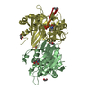
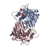
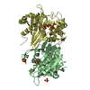
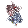
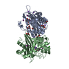
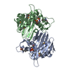
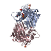
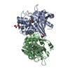
| Entry | Database: PDB / ID: 5fq9 | ||||||
|---|---|---|---|---|---|---|---|
| Title | Crystal structure of the OXA10 with 1C | ||||||
 Components Components | BETA-LACTAMASE OXA-10 | ||||||
 Keywords Keywords | HYDROLASE / ANTIBIOTIC RESISTANCE | ||||||
| Function / homology |  Function and homology information Function and homology informationpenicillin binding / antibiotic catabolic process / cell wall organization / beta-lactamase activity / beta-lactamase / periplasmic space / response to antibiotic / plasma membrane Similarity search - Function | ||||||
| Biological species |   PSEUDOMONAS AERUGINOSA (bacteria) PSEUDOMONAS AERUGINOSA (bacteria) | ||||||
| Method |  X-RAY DIFFRACTION / SYNCHROTRON / MOLECULAR REPLACEMENT / Resolution: 1.5 Å X-RAY DIFFRACTION / SYNCHROTRON / MOLECULAR REPLACEMENT / Resolution: 1.5 Å | ||||||
 Authors Authors | Brem, J. / McDonough, M.A. / Cain, R. / Clifton, I. / Fishwick, C.W.G. / Schofield, C.J. | ||||||
 Citation Citation | Journal: Nat Commun / Year: 2016 Title: Structural basis of metallo-beta-lactamase, serine-beta-lactamase and penicillin-binding protein inhibition by cyclic boronates. Authors: Brem, J. / Cain, R. / Cahill, S. / McDonough, M.A. / Clifton, I.J. / Jimenez-Castellanos, J.C. / Avison, M.B. / Spencer, J. / Fishwick, C.W. / Schofield, C.J. | ||||||
| History |
|
- Structure visualization
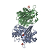
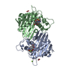
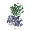
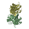
| Structure viewer | Molecule: MolmilJmol/JSmol |
|---|
- Downloads & links
-Download
| PDBx/mmCIF format | 5fq9.cif.gz | 220.1 KB | Display | PDBx/mmCIF format |
|---|---|---|---|---|
| PDB format | pdb5fq9.ent.gz | 176.4 KB | Display | PDB format |
| PDBx/mmJSON format | 5fq9.json.gz | Tree view | PDBx/mmJSON format | |
| Others |  Other downloads Other downloads |
-Validation report
| Arichive directory | https://data.pdbj.org/pub/pdb/validation_reports/fq/5fq9ftp://data.pdbj.org/pub/pdb/validation_reports/fq/5fq9 | HTTPS FTP |
|---|
-Related structure data
| Related structure data | 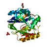 5fqbC  5fqcC  5j8xC  1k6rS C: citing same article ( S: Starting model for refinement |
|---|---|
| Similar structure data |


-Links
 PDBj
PDBj- Assembly
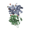
| Deposited unit | 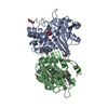
| ||||||||
|---|---|---|---|---|---|---|---|---|---|
| 1 |
| ||||||||
| Unit cell |
| ||||||||
| Noncrystallographic symmetry (NCS) | NCS oper: (Code: given Matrix: (0.8054, 0.591, 0.0452), Vector: |
-Components
-Protein , 1 types, 2 molecules AB
| #1: Protein | Mass: 27755.541 Da / Num. of mol.: 2 Source method: isolated from a genetically manipulated source Details: 1C BOUND TO THE ACTIVE SITE / Source: (gene. exp.) PSEUDOMONAS AERUGINOSA (bacteria) / Production host: |
|---|
-Non-polymers , 6 types, 500 molecules 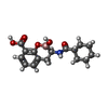


| #2: Chemical |  Mass: 317.145 Da / Num. of mol.: 2 / Source method: obtained synthetically / Formula: C16H20BNO5 Mass: 317.145 Da / Num. of mol.: 2 / Source method: obtained synthetically / Formula: C16H20BNO5#3: Chemical | ChemComp-GOL /  Mass: 92.094 Da / Num. of mol.: 4 / Source method: obtained synthetically / Formula: C3H8O3 Mass: 92.094 Da / Num. of mol.: 4 / Source method: obtained synthetically / Formula: C3H8O3#4: Chemical | ChemComp-ACT / |  Mass: 59.044 Da / Num. of mol.: 1 / Source method: obtained synthetically / Formula: C2H3O2 Mass: 59.044 Da / Num. of mol.: 1 / Source method: obtained synthetically / Formula: C2H3O2#5: Chemical | ChemComp-CL / |  Mass: 35.453 Da / Num. of mol.: 1 / Source method: obtained synthetically / Formula: Cl Mass: 35.453 Da / Num. of mol.: 1 / Source method: obtained synthetically / Formula: Cl#6: Chemical | ChemComp-NA / |  Mass: 22.990 Da / Num. of mol.: 1 / Source method: obtained synthetically / Formula: Na Mass: 22.990 Da / Num. of mol.: 1 / Source method: obtained synthetically / Formula: Na#7: Water | ChemComp-HOH / | Mass: 18.015 Da / Num. of mol.: 491 / Source method: isolated from a natural source / Formula: H2O |
|---|
-Experimental details
-Experiment
| Experiment | Method: X-RAY DIFFRACTION / Number of used crystals: 1 |
|---|
- Sample preparation
| Crystal | Density Matthews: 3.17 Å3/Da / Density % sol: 61.15 % / Description: NONE |
|---|---|
| Crystal grow | pH: 5.5 / Details: 0.2M NACL, 0.1M NA ACETATE PH 5.0, 20% PEG 6000 |
-Data collection
| Diffraction | Mean temperature: 100 K |
|---|---|
| Diffraction source | Source: SYNCHROTRON / Site: Diamond  / Beamline: I04 / Wavelength: 0.9795 / Beamline: I04 / Wavelength: 0.9795 |
| Detector | Type: DECTRIS PILATUS 6M / Detector: PIXEL / Date: Sep 27, 2015 / Details: MIRRORS |
| Radiation | Monochromator: DOUBLE CRYSTAL / Protocol: SINGLE WAVELENGTH / Monochromatic (M) / Laue (L): M / Scattering type: x-ray |
| Radiation wavelength | Wavelength: 0.9795 Å / Relative weight: 1 |
| Reflection | Resolution: 1.5→15.44 Å / Num. obs: 253261 / % possible obs: 99.9 % / Observed criterion σ(I): 2 / Redundancy: 5.9 % / Biso Wilson estimate: 16.1 Å2 / Rmerge(I) obs: 0.05 / Net I/σ(I): 5.6 |
| Reflection shell | Resolution: 1.1→1.12 Å / Redundancy: 8.9 % / Rmerge(I) obs: 1.1 / Mean I/σ(I) obs: 2 / % possible all: 99.5 |
- Processing
| Software |
| |||||||||||||||||||||||||||||||||||||||||||||||||||||||||||||||||||||||||||||||||||||||||||||||||||||||||||||||||||||||||||||||||||||||||||||||||||||||||||||||||||||||||||||||||||||||||||||||||||||||||||||||||||||||||
|---|---|---|---|---|---|---|---|---|---|---|---|---|---|---|---|---|---|---|---|---|---|---|---|---|---|---|---|---|---|---|---|---|---|---|---|---|---|---|---|---|---|---|---|---|---|---|---|---|---|---|---|---|---|---|---|---|---|---|---|---|---|---|---|---|---|---|---|---|---|---|---|---|---|---|---|---|---|---|---|---|---|---|---|---|---|---|---|---|---|---|---|---|---|---|---|---|---|---|---|---|---|---|---|---|---|---|---|---|---|---|---|---|---|---|---|---|---|---|---|---|---|---|---|---|---|---|---|---|---|---|---|---|---|---|---|---|---|---|---|---|---|---|---|---|---|---|---|---|---|---|---|---|---|---|---|---|---|---|---|---|---|---|---|---|---|---|---|---|---|---|---|---|---|---|---|---|---|---|---|---|---|---|---|---|---|---|---|---|---|---|---|---|---|---|---|---|---|---|---|---|---|---|---|---|---|---|---|---|---|---|---|---|---|---|---|---|---|---|
| Refinement | Method to determine structure: MOLECULAR REPLACEMENT Starting model: PDB ENTRY 1K6R Resolution: 1.5→15.424 Å / SU ML: 0.15 / σ(F): 1.34 / Phase error: 17.79 / Stereochemistry target values: ML
| |||||||||||||||||||||||||||||||||||||||||||||||||||||||||||||||||||||||||||||||||||||||||||||||||||||||||||||||||||||||||||||||||||||||||||||||||||||||||||||||||||||||||||||||||||||||||||||||||||||||||||||||||||||||||
| Solvent computation | Shrinkage radii: 0.9 Å / VDW probe radii: 1.11 Å / Solvent model: FLAT BULK SOLVENT MODEL | |||||||||||||||||||||||||||||||||||||||||||||||||||||||||||||||||||||||||||||||||||||||||||||||||||||||||||||||||||||||||||||||||||||||||||||||||||||||||||||||||||||||||||||||||||||||||||||||||||||||||||||||||||||||||
| Displacement parameters | Biso mean: 16.325 Å2 | |||||||||||||||||||||||||||||||||||||||||||||||||||||||||||||||||||||||||||||||||||||||||||||||||||||||||||||||||||||||||||||||||||||||||||||||||||||||||||||||||||||||||||||||||||||||||||||||||||||||||||||||||||||||||
| Refinement step | Cycle: LAST / Resolution: 1.5→15.424 Å
| |||||||||||||||||||||||||||||||||||||||||||||||||||||||||||||||||||||||||||||||||||||||||||||||||||||||||||||||||||||||||||||||||||||||||||||||||||||||||||||||||||||||||||||||||||||||||||||||||||||||||||||||||||||||||
| Refine LS restraints |
| |||||||||||||||||||||||||||||||||||||||||||||||||||||||||||||||||||||||||||||||||||||||||||||||||||||||||||||||||||||||||||||||||||||||||||||||||||||||||||||||||||||||||||||||||||||||||||||||||||||||||||||||||||||||||
| LS refinement shell |
|
