 Movie
Movie Controller


+ Open data

- Basic information
| Entry | Database: PDB / ID: 1e4d | ||||||
|---|---|---|---|---|---|---|---|
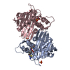
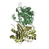
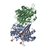
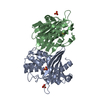
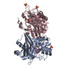
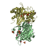
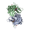
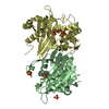
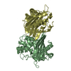
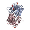
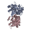
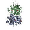
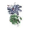
| Title | Structure of OXA10 beta-lactamase at pH 8.3 | ||||||
 Components Components | BETA-LACTAMASE OXA-10 | ||||||
 Keywords Keywords | BETA-LACTAMASE / ANTIOBITIC RESISTANCE | ||||||
| Function / homology |  Function and homology information Function and homology informationpenicillin binding / antibiotic catabolic process / cell wall organization / beta-lactamase activity / beta-lactamase / periplasmic space / response to antibiotic / plasma membrane Similarity search - Function | ||||||
| Biological species |   PSEUDOMONAS AERUGINOSA (bacteria) PSEUDOMONAS AERUGINOSA (bacteria) | ||||||
| Method |  X-RAY DIFFRACTION / SYNCHROTRON / MOLECULAR REPLACEMENT / Resolution: 1.8 Å X-RAY DIFFRACTION / SYNCHROTRON / MOLECULAR REPLACEMENT / Resolution: 1.8 Å | ||||||
 Authors Authors | Maveyraud, L. / Golemi, D. / Kotra, L.P. / Tranier, S. / Vakulenko, S. / Mobashery, S. / Samama, J.P. | ||||||
 Citation Citation | Journal: Structure / Year: 2000 Title: Insights Into Class D Beta-Lactamases are Revealed by the Crystal Structure of the Oxa10 Enzyme from Pseudomonas Aeruginosa Authors: Maveyraud, L. / Golemi, D. / Kotra, L.P. / Tranier, S. / Vakulenko, S. / Mobashery, S. / Samama, J.P. | ||||||
| History |
|
- Structure visualization
| Structure viewer | Molecule: MolmilJmol/JSmol |
|---|
- Downloads & links
-Download
| PDBx/mmCIF format | 1e4d.cif.gz | 417.1 KB | Display | PDBx/mmCIF format |
|---|---|---|---|---|
| PDB format | pdb1e4d.ent.gz | 342.2 KB | Display | PDB format |
| PDBx/mmJSON format | 1e4d.json.gz | Tree view | PDBx/mmJSON format | |
| Others |  Other downloads Other downloads |
-Validation report
| Arichive directory | https://data.pdbj.org/pub/pdb/validation_reports/e4/1e4dftp://data.pdbj.org/pub/pdb/validation_reports/e4/1e4d | HTTPS FTP |
|---|
-Related structure data
| Related structure data | 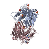 1e3uSC S: Starting model for refinement C: citing same article ( |
|---|---|
| Similar structure data |


-Links
 PDBj
PDBj- Assembly
| Deposited unit | 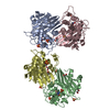
| ||||||||||||||||
|---|---|---|---|---|---|---|---|---|---|---|---|---|---|---|---|---|---|
| 1 | 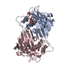
| ||||||||||||||||
| 2 | 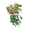
| ||||||||||||||||
| Unit cell |
| ||||||||||||||||
| Noncrystallographic symmetry (NCS) | NCS oper:
|
-Components
| #1: Protein | Mass: 27567.293 Da / Num. of mol.: 4 Source method: isolated from a genetically manipulated source Source: (gene. exp.) PSEUDOMONAS AERUGINOSA (bacteria) / Cellular location (production host): EXCRETED / Production host: #2: Chemical | ChemComp-SO4 /   Mass: 96.063 Da / Num. of mol.: 16 / Source method: obtained synthetically / Formula: SO4 Mass: 96.063 Da / Num. of mol.: 16 / Source method: obtained synthetically / Formula: SO4#3: Chemical | ChemComp-EDO /   Mass: 62.068 Da / Num. of mol.: 6 / Source method: obtained synthetically / Formula: C2H6O2 Mass: 62.068 Da / Num. of mol.: 6 / Source method: obtained synthetically / Formula: C2H6O2#4: Water | ChemComp-HOH / |  Mass: 18.015 Da / Num. of mol.: 943 / Source method: isolated from a natural source / Formula: H2O Mass: 18.015 Da / Num. of mol.: 943 / Source method: isolated from a natural source / Formula: H2O |
|---|
-Experimental details
-Experiment
| Experiment | Method: X-RAY DIFFRACTION / Number of used crystals: 1 |
|---|
- Sample preparation
| Crystal | Density Matthews: 2.53 Å3/Da / Density % sol: 0.561 % | |||||||||||||||||||||||||
|---|---|---|---|---|---|---|---|---|---|---|---|---|---|---|---|---|---|---|---|---|---|---|---|---|---|---|
| Crystal grow | pH: 8.3 Details: AMMONIUMS SULFATE 2.0 M, TRIS HCL 100 MM, PH 8.2-8.5 | |||||||||||||||||||||||||
| Crystal grow | *PLUS Temperature: 4 ℃ / pH: 7.8 / Method: vapor diffusion, hanging drop | |||||||||||||||||||||||||
| Components of the solutions | *PLUS
|
-Data collection
| Diffraction | Mean temperature: 100 K |
|---|---|
| Diffraction source | Source: SYNCHROTRON / Site: ESRF  / Beamline: BM30A / Wavelength: 0.9796 / Beamline: BM30A / Wavelength: 0.9796 |
| Detector | Type: MARRESEARCH / Detector: IMAGE PLATE / Date: Feb 15, 2000 |
| Radiation | Protocol: SINGLE WAVELENGTH / Monochromatic (M) / Laue (L): M / Scattering type: x-ray |
| Radiation wavelength | Wavelength: 0.9796 Å / Relative weight: 1 |
| Reflection | Resolution: 1.8→29.54 Å / Num. obs: 86153 / % possible obs: 84.4 % / Redundancy: 3 % / Biso Wilson estimate: 22.19 Å2 / Rsym value: 0.086 / Net I/σ(I): 12.3 |
| Reflection shell | Resolution: 1.8→1.89 Å / Redundancy: 2.6 % / Mean I/σ(I) obs: 2.7 / Rsym value: 0.331 / % possible all: 90.7 |
| Reflection | *PLUS Num. measured all: 258171 / Rmerge(I) obs: 0.086 |
| Reflection shell | *PLUS % possible obs: 91.1 % / Rmerge(I) obs: 0.331 |
- Processing
| Software |
| ||||||||||||||||||||||||||||||||||||||||||||||||||||||||||||||||||||||||||||||||||||
|---|---|---|---|---|---|---|---|---|---|---|---|---|---|---|---|---|---|---|---|---|---|---|---|---|---|---|---|---|---|---|---|---|---|---|---|---|---|---|---|---|---|---|---|---|---|---|---|---|---|---|---|---|---|---|---|---|---|---|---|---|---|---|---|---|---|---|---|---|---|---|---|---|---|---|---|---|---|---|---|---|---|---|---|---|---|
| Refinement | Method to determine structure: MOLECULAR REPLACEMENT Starting model: 1E3U Resolution: 1.8→29.54 Å / SU B: 1.95692 / SU ML: 0.06092 / Cross valid method: THROUGHOUT / σ(F): 0 / ESU R: 0.86904 / ESU R Free: 0.15506 Details: INDIVIDUAL ANISOTROPIC BFACTORS WERE REFINED. THE C-TERMINAL RESIDUE WAS NOT SEEN IN THE DENSITY MAPS
| ||||||||||||||||||||||||||||||||||||||||||||||||||||||||||||||||||||||||||||||||||||
| Displacement parameters | Biso mean: 22.35 Å2 | ||||||||||||||||||||||||||||||||||||||||||||||||||||||||||||||||||||||||||||||||||||
| Refinement step | Cycle: LAST / Resolution: 1.8→29.54 Å
| ||||||||||||||||||||||||||||||||||||||||||||||||||||||||||||||||||||||||||||||||||||
| Refine LS restraints |
| ||||||||||||||||||||||||||||||||||||||||||||||||||||||||||||||||||||||||||||||||||||
| Software | *PLUS Name: REFMAC / Classification: refinement | ||||||||||||||||||||||||||||||||||||||||||||||||||||||||||||||||||||||||||||||||||||
| Refinement | *PLUS Rfactor obs: 0.1983 / Rfactor Rfree: 0.24271 / Rfactor Rwork: 0.19831 | ||||||||||||||||||||||||||||||||||||||||||||||||||||||||||||||||||||||||||||||||||||
| Solvent computation | *PLUS | ||||||||||||||||||||||||||||||||||||||||||||||||||||||||||||||||||||||||||||||||||||
| Displacement parameters | *PLUS |
