 Movie
Movie Controller

[English] 日本語
 Yorodumi
- PDB-6skq: OXA-655_MEM. Structural insights into the enhanced carbapenemase ... -
+ Open data

- Basic information
| Entry | Database: PDB / ID: 6skq | ||||||
|---|---|---|---|---|---|---|---|
| Title | OXA-655_MEM. Structural insights into the enhanced carbapenemase efficiency of OXA-655 compared to OXA-10. | ||||||
 Components Components | Beta-lactamase | ||||||
 Keywords Keywords | ANTIBIOTIC / Class D beta-lactamase / crystal structures / Carbapenemases / Antibiotic resistance | ||||||
| Function / homology |  Function and homology information Function and homology informationpenicillin binding / antibiotic catabolic process / cell wall organization / beta-lactamase activity / beta-lactamase / response to antibiotic Similarity search - Function | ||||||
| Biological species |  | ||||||
| Method |  X-RAY DIFFRACTION / SYNCHROTRON / MOLECULAR REPLACEMENT / Resolution: 2.1 Å X-RAY DIFFRACTION / SYNCHROTRON / MOLECULAR REPLACEMENT / Resolution: 2.1 Å | ||||||
 Authors Authors | Leiros, H.-K.S. | ||||||
 Citation Citation | Journal: Febs Open Bio / Year: 2020 Title: Structural insights into the enhanced carbapenemase efficiency of OXA-655 compared to OXA-10. Authors: Leiros, H.S. / Thomassen, A.M. / Samuelsen, O. / Flach, C.F. / Kotsakis, S.D. / Larsson, D.G.J. | ||||||
| History |
|
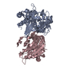
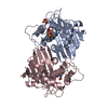
- Structure visualization
| Structure viewer | Molecule: MolmilJmol/JSmol |
|---|
- Downloads & links
-Download
| PDBx/mmCIF format | 6skq.cif.gz | 491.5 KB | Display | PDBx/mmCIF format |
|---|---|---|---|---|
| PDB format | pdb6skq.ent.gz | 330.5 KB | Display | PDB format |
| PDBx/mmJSON format | 6skq.json.gz | Tree view | PDBx/mmJSON format | |
| Others |  Other downloads Other downloads |
-Validation report
| Arichive directory | https://data.pdbj.org/pub/pdb/validation_reports/sk/6skqftp://data.pdbj.org/pub/pdb/validation_reports/sk/6skq | HTTPS FTP |
|---|
-Related structure data
| Related structure data |  6skpC  6skrC 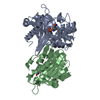 4s2oS C: citing same article ( S: Starting model for refinement |
|---|---|
| Similar structure data |


-Links
 PDBj
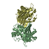
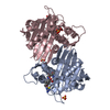
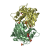
PDBj- Assembly
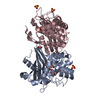
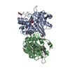
| Deposited unit | 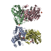
| ||||||||||||
|---|---|---|---|---|---|---|---|---|---|---|---|---|---|
| 1 | 
| ||||||||||||
| 2 | 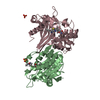
| ||||||||||||
| Unit cell |
|
-Components
| #1: Protein | Mass: 29625.836 Da / Num. of mol.: 4 Source method: isolated from a genetically manipulated source Source: (gene. exp.) #2: Chemical | ChemComp-MER / (   Mass: 385.478 Da / Num. of mol.: 4 / Source method: obtained synthetically / Formula: C17H27N3O5S / Feature type: SUBJECT OF INVESTIGATION / Comment: antibiotic*YM Mass: 385.478 Da / Num. of mol.: 4 / Source method: obtained synthetically / Formula: C17H27N3O5S / Feature type: SUBJECT OF INVESTIGATION / Comment: antibiotic*YM#3: Chemical | ChemComp-SO4 /   Mass: 96.063 Da / Num. of mol.: 5 / Source method: obtained synthetically / Formula: SO4 Mass: 96.063 Da / Num. of mol.: 5 / Source method: obtained synthetically / Formula: SO4#4: Water | ChemComp-HOH / |  Mass: 18.015 Da / Num. of mol.: 970 / Source method: isolated from a natural source / Formula: H2O Mass: 18.015 Da / Num. of mol.: 970 / Source method: isolated from a natural source / Formula: H2OHas ligand of interest | Y | |
|---|
-Experimental details
-Experiment
| Experiment | Method: X-RAY DIFFRACTION / Number of used crystals: 1 |
|---|
- Sample preparation
| Crystal | Density Matthews: 2.5 Å3/Da / Density % sol: 50.88 % |
|---|---|
| Crystal grow | Temperature: 277 K / Method: vapor diffusion, hanging drop / Details: 6.8 mg/mL, 20-23% PEG 3350 0.2 M LiSO4 |
-Data collection
| Diffraction | Mean temperature: 100 K / Serial crystal experiment: N |
|---|---|
| Diffraction source | Source: SYNCHROTRON / Site: BESSY  / Beamline: 14.1 / Wavelength: 0.9184 Å / Beamline: 14.1 / Wavelength: 0.9184 Å |
| Detector | Type: PSI PILATUS 6M / Detector: PIXEL / Date: Feb 18, 2018 |
| Radiation | Protocol: SINGLE WAVELENGTH / Monochromatic (M) / Laue (L): M / Scattering type: x-ray |
| Radiation wavelength | Wavelength: 0.9184 Å / Relative weight: 1 |
| Reflection | Resolution: 2.1→25 Å / Num. obs: 61827 / % possible obs: 94.9 % / Redundancy: 5.8 % / Biso Wilson estimate: 18.34 Å2 / CC1/2: 0.979 / Rrim(I) all: 0.185 / Net I/σ(I): 5.8 |
| Reflection shell | Resolution: 2.1→2.15 Å / Num. unique obs: 4483 / CC1/2: 0.539 / Rrim(I) all: 0.824 |
- Processing
| Software |
| ||||||||||||||||||||||||||||||||||||||||||||||||||||||||||||||||||||||||||||||||||||||||||||||||||||||||||||||||||||||||||||||
|---|---|---|---|---|---|---|---|---|---|---|---|---|---|---|---|---|---|---|---|---|---|---|---|---|---|---|---|---|---|---|---|---|---|---|---|---|---|---|---|---|---|---|---|---|---|---|---|---|---|---|---|---|---|---|---|---|---|---|---|---|---|---|---|---|---|---|---|---|---|---|---|---|---|---|---|---|---|---|---|---|---|---|---|---|---|---|---|---|---|---|---|---|---|---|---|---|---|---|---|---|---|---|---|---|---|---|---|---|---|---|---|---|---|---|---|---|---|---|---|---|---|---|---|---|---|---|---|
| Refinement | Method to determine structure: MOLECULAR REPLACEMENT Starting model: 4s2o Resolution: 2.1→24.05 Å / SU ML: 0.2467 / Cross valid method: THROUGHOUT / σ(F): 1.34 / Phase error: 23.5815
| ||||||||||||||||||||||||||||||||||||||||||||||||||||||||||||||||||||||||||||||||||||||||||||||||||||||||||||||||||||||||||||||
| Solvent computation | Shrinkage radii: 0.9 Å / VDW probe radii: 1.11 Å | ||||||||||||||||||||||||||||||||||||||||||||||||||||||||||||||||||||||||||||||||||||||||||||||||||||||||||||||||||||||||||||||
| Displacement parameters | Biso mean: 21.26 Å2 | ||||||||||||||||||||||||||||||||||||||||||||||||||||||||||||||||||||||||||||||||||||||||||||||||||||||||||||||||||||||||||||||
| Refinement step | Cycle: LAST / Resolution: 2.1→24.05 Å
| ||||||||||||||||||||||||||||||||||||||||||||||||||||||||||||||||||||||||||||||||||||||||||||||||||||||||||||||||||||||||||||||
| Refine LS restraints |
| ||||||||||||||||||||||||||||||||||||||||||||||||||||||||||||||||||||||||||||||||||||||||||||||||||||||||||||||||||||||||||||||
| LS refinement shell |
|
