 Movie
Movie Controller


+ Open data

- Basic information
| Entry | Database: PDB / ID: 1k55 | ||||||
|---|---|---|---|---|---|---|---|
| Title | OXA 10 class D beta-lactamase at pH 7.5 | ||||||
 Components Components | (Beta lactamase OXA-10) x 2 | ||||||
 Keywords Keywords | HYDROLASE / beta-lactamase / antibiotic resistance / carbamylation | ||||||
| Function / homology |  Function and homology information Function and homology informationpenicillin binding / antibiotic catabolic process / cell wall organization / beta-lactamase activity / beta-lactamase / periplasmic space / response to antibiotic / plasma membrane Similarity search - Function | ||||||
| Biological species |   Pseudomonas aeruginosa (bacteria) Pseudomonas aeruginosa (bacteria) | ||||||
| Method |  X-RAY DIFFRACTION / SYNCHROTRON / FOURIER SYNTHESIS / Resolution: 1.39 Å X-RAY DIFFRACTION / SYNCHROTRON / FOURIER SYNTHESIS / Resolution: 1.39 Å | ||||||
 Authors Authors | Golemi, D. / Maveyraud, L. / Vakulenko, S. / Samama, J.P. / Mobashery, S. | ||||||
 Citation Citation | Journal: Proc.Natl.Acad.Sci.USA / Year: 2001 Title: Critical involvement of a carbamylated lysine in catalytic function of class D beta-lactamases. Authors: Golemi, D. / Maveyraud, L. / Vakulenko, S. / Samama, J.P. / Mobashery, S. #1: Journal: Structure / Year: 2000Title: Insights into class D beta-lactamases are revealed by the crystal structure of the OXA10 enzyme from Pseudomonas aeruginosa Authors: Maveyraud, L. / Golemi, D. / Kotra, L.P. / Tranier, S. / Vakulenko, S. / Mobashery, S. / Samama, J.P. #2: Journal: J.Am.Chem.Soc. / Year: 2000Title: The first structural and mechanistic insights for class D beta-lactamases: evidence for a novel catalytic process for turnover of beta-lactam antibiotic Authors: Golemi, D. / Maveyraud, L. / Vakulenko, S. / Tranier, S. / Ishiwata, A. / Kotra, L.P. / Samama, J.P. / Mobashery, S. | ||||||
| History |
|
- Structure visualization
| Structure viewer | Molecule: MolmilJmol/JSmol |
|---|
- Downloads & links
-Download
| PDBx/mmCIF format | 1k55.cif.gz | 437.3 KB | Display | PDBx/mmCIF format |
|---|---|---|---|---|
| PDB format | pdb1k55.ent.gz | 357.4 KB | Display | PDB format |
| PDBx/mmJSON format | 1k55.json.gz | Tree view | PDBx/mmJSON format | |
| Others |  Other downloads Other downloads |
-Validation report
| Arichive directory | https://data.pdbj.org/pub/pdb/validation_reports/k5/1k55ftp://data.pdbj.org/pub/pdb/validation_reports/k5/1k55 | HTTPS FTP |
|---|
-Related structure data
| Related structure data |  1k54C  1k56C 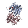 1k57C 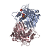 1e4dS S: Starting model for refinement C: citing same article ( |
|---|---|
| Similar structure data |


-Links
 PDBj
PDBj- Assembly
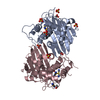
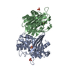
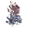
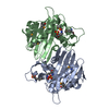
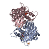
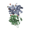
| Deposited unit | 
| ||||||||
|---|---|---|---|---|---|---|---|---|---|
| 1 | 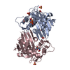
| ||||||||
| 2 | 
| ||||||||
| 3 | 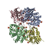
| ||||||||
| Unit cell |
| ||||||||
| Details | the biological assembly is a dimer. There are two dimers in the asymmetric unit : chains A and C form a dimer chains B and D form a dimer |
-Components
| #1: Protein | Mass: 27567.293 Da / Num. of mol.: 2 Source method: isolated from a genetically manipulated source Details: RESIDUE 70 OF CHAINS A AND B, KCX ARE CARBAMYLATED LYSINE. Source: (gene. exp.) Pseudomonas aeruginosa (bacteria) / Plasmid: pET24a / Species (production host): Escherichia coli / Production host: #2: Protein | Mass: 27524.291 Da / Num. of mol.: 2 Source method: isolated from a genetically manipulated source Details: RESIDUE 70 OF CHAINS C AND D EXISTS IN TWO ALTERNATE CONFORMATIONS: KCX, CARBAMYLATED LYSINE, AND LYSINE. Source: (gene. exp.) Pseudomonas aeruginosa (bacteria) / Plasmid: pET24a / Species (production host): Escherichia coli / Production host: #3: Chemical | ChemComp-SO4 /   Mass: 96.063 Da / Num. of mol.: 18 / Source method: obtained synthetically / Formula: SO4 Mass: 96.063 Da / Num. of mol.: 18 / Source method: obtained synthetically / Formula: SO4#4: Chemical | ChemComp-EDO /   Mass: 62.068 Da / Num. of mol.: 9 / Source method: obtained synthetically / Formula: C2H6O2 Mass: 62.068 Da / Num. of mol.: 9 / Source method: obtained synthetically / Formula: C2H6O2#5: Water | ChemComp-HOH / |  Mass: 18.015 Da / Num. of mol.: 942 / Source method: isolated from a natural source / Formula: H2O Mass: 18.015 Da / Num. of mol.: 942 / Source method: isolated from a natural source / Formula: H2OHas protein modification | Y | |
|---|
-Experimental details
-Experiment
| Experiment | Method: X-RAY DIFFRACTION / Number of used crystals: 1 |
|---|
- Sample preparation
| Crystal | Density Matthews: 2.51 Å3/Da / Density % sol: 51.04 % | ||||||||||||||||||||||||||||||
|---|---|---|---|---|---|---|---|---|---|---|---|---|---|---|---|---|---|---|---|---|---|---|---|---|---|---|---|---|---|---|---|
| Crystal grow | Temperature: 277 K / Method: vapor diffusion, hanging drop / pH: 7.5 Details: ammonium sulphate, HEPES, pH 7.5, VAPOR DIFFUSION, HANGING DROP, temperature 277K | ||||||||||||||||||||||||||||||
| Crystal grow | *PLUS Temperature: 4 ℃ / pH: 7.8 | ||||||||||||||||||||||||||||||
| Components of the solutions | *PLUS
|
-Data collection
| Diffraction | Mean temperature: 100 K |
|---|---|
| Diffraction source | Source: SYNCHROTRON / Site: ESRF  / Beamline: ID14-1 / Wavelength: 0.934 Å / Beamline: ID14-1 / Wavelength: 0.934 Å |
| Detector | Type: MARRESEARCH / Detector: CCD / Date: Jun 6, 2000 |
| Radiation | Monochromator: Germanium Ge(220) / Protocol: SINGLE WAVELENGTH / Monochromatic (M) / Laue (L): M / Scattering type: x-ray |
| Radiation wavelength | Wavelength: 0.934 Å / Relative weight: 1 |
| Reflection | Resolution: 1.39→53.16 Å / Num. all: 216019 / Num. obs: 216019 / % possible obs: 99 % / Observed criterion σ(F): 0 / Observed criterion σ(I): 0 / Redundancy: 3.9 % / Biso Wilson estimate: 16.4 Å2 / Rmerge(I) obs: 0.082 / Rsym value: 0.082 / Net I/σ(I): 11.8 |
| Reflection shell | Resolution: 1.39→1.47 Å / Redundancy: 3.2 % / Rmerge(I) obs: 0.373 / Mean I/σ(I) obs: 3.3 / Num. unique all: 29650 / Rsym value: 0.373 / % possible all: 99 |
| Reflection | *PLUS Num. measured all: 841487 |
| Reflection shell | *PLUS % possible obs: 93.3 % / Num. unique obs: 29650 / Num. measured obs: 95038 |
- Processing
| Software |
| |||||||||||||||||||||||||
|---|---|---|---|---|---|---|---|---|---|---|---|---|---|---|---|---|---|---|---|---|---|---|---|---|---|---|
| Refinement | Method to determine structure: FOURIER SYNTHESIS Starting model: PDB entry 1E4D Resolution: 1.39→53.16 Å / Isotropic thermal model: overall anisotropic / Cross valid method: THROUGHOUT / σ(F): 0 / Stereochemistry target values: engh & huber Details: SUL 1024, which is assigned alternate position B, corresponds to alternate conformation B of chain D residue 115. Chain C: the following residues display alternate conformations for side ...Details: SUL 1024, which is assigned alternate position B, corresponds to alternate conformation B of chain D residue 115. Chain C: the following residues display alternate conformations for side chain atoms only : Thr23C S60C, Glu195C, Glu231C and Ser245C. Chain D: the following residues display alternate conformations for side chain atoms only : Ser33C Ser60D, Glu86D, Arg125D, Ser147D, Ser179D, Glu183D. Gly265D and Gly266D display alternate conformations of the dipeptide. chains C and D: Other residues in alternate conformation correspond to two distinct conformations of the enzyme: conformations A and B : Met99C, Lys100C, Gln101C, Trp102C, Glu103C, Val114C, Ser115C, Ala116C, Val117C and Pro118C. RESIDUE 70 EXISTS IN TWO CONFORMATIONS: CONFORMATION A, AS a classical non modified LYS residue, AND CONFORMATION B, AS KCX, a carbamylated lysine,in chains C and D. There is no density accounting for conformation B of residues Arg97 C and D, Ala98 C and D and Met99D, but these residues are necessarly in an alternate conformation for sterical reasons.
| |||||||||||||||||||||||||
| Displacement parameters | Biso mean: 14.97 Å2
| |||||||||||||||||||||||||
| Refinement step | Cycle: LAST / Resolution: 1.39→53.16 Å
| |||||||||||||||||||||||||
| Refine LS restraints |
| |||||||||||||||||||||||||
| LS refinement shell | Resolution: 1.39→1.427 Å
| |||||||||||||||||||||||||
| Software | *PLUS Name: REFMAC / Classification: refinement | |||||||||||||||||||||||||
| Refinement | *PLUS σ(F): 0 / % reflection Rfree: 1 % / Rfactor obs: 0.15311 / Rfactor Rfree: 0.1809 | |||||||||||||||||||||||||
| Solvent computation | *PLUS | |||||||||||||||||||||||||
| Displacement parameters | *PLUS | |||||||||||||||||||||||||
| Refine LS restraints | *PLUS
|
