 Movie
Movie Controller

[English] 日本語
 Yorodumi
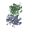
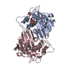
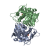
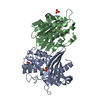
- PDB-1h8y: Crystal structure of the class D beta-lactamase OXA-13 in complex... -
+ Open data

- Basic information
| Entry | Database: PDB / ID: 1h8y | ||||||
|---|---|---|---|---|---|---|---|
| Title | Crystal structure of the class D beta-lactamase OXA-13 in complex with meropenem | ||||||
 Components Components | BETA-LACTAMASE | ||||||
 Keywords Keywords | HYDROLASE / BETA-LACTAMASE / CLASS D / OXA-13 / MEROPENEM / ACYL- ENZYME | ||||||
| Function / homology |  Function and homology information Function and homology informationpenicillin binding / antibiotic catabolic process / cell wall organization / beta-lactamase activity / beta-lactamase / response to antibiotic / plasma membrane Similarity search - Function | ||||||
| Biological species |   PSEUDOMONAS AERUGINOSA (bacteria) PSEUDOMONAS AERUGINOSA (bacteria) | ||||||
| Method |  X-RAY DIFFRACTION / SYNCHROTRON / MOLECULAR REPLACEMENT / Resolution: 2 Å X-RAY DIFFRACTION / SYNCHROTRON / MOLECULAR REPLACEMENT / Resolution: 2 Å | ||||||
 Authors Authors | Pernot, L. / Frenois, F. / Rybkine, T. / L'Hermite, G. / Petrella, S. / Delettre, J. / Jarlier, V. / Collatz, E. / Sougakoff, W. | ||||||
 Citation Citation | Journal: J.Mol.Biol. / Year: 2001 Title: Crystal Structures of the Class D B-Lactamase Oxa-13 in the Native Form and in Complex with Meropenem Authors: Pernot, L. / Frenois, F. / Rybkine, T. / L'Hermite, G. / Petrella, S. / Delettre, J. / Jarlier, V. / Collatz, E. / Sougakoff, W. #1: Journal: Microbiology / Year: 1998 Title: Carbapenemns as Inhibitors of Oxa-13, a Novel, Integron-Encoded B-Lactmase in Pseudomonas Aeruginosa Authors: Mugnier, P. / Podglajen, I. / Goldstein, F.W. / Collatz, E. | ||||||
| History |
| ||||||
| Remark 650 | HELIX DETERMINATION METHOD: AUTHOR PROVIDED. | ||||||
| Remark 700 | SHEET DETERMINATION METHOD: AUTHOR PROVIDED. |
- Structure visualization
| Structure viewer | Molecule: MolmilJmol/JSmol |
|---|
- Downloads & links
-Download
| PDBx/mmCIF format | 1h8y.cif.gz | 117.7 KB | Display | PDBx/mmCIF format |
|---|---|---|---|---|
| PDB format | pdb1h8y.ent.gz | 91.8 KB | Display | PDB format |
| PDBx/mmJSON format | 1h8y.json.gz | Tree view | PDBx/mmJSON format | |
| Others |  Other downloads Other downloads |
-Validation report
| Arichive directory | https://data.pdbj.org/pub/pdb/validation_reports/h8/1h8yftp://data.pdbj.org/pub/pdb/validation_reports/h8/1h8y | HTTPS FTP |
|---|
-Related structure data


-Links
 PDBj
PDBj- Assembly
| Deposited unit | 
| ||||||||
|---|---|---|---|---|---|---|---|---|---|
| 1 |
| ||||||||
| Unit cell |
| ||||||||
| Noncrystallographic symmetry (NCS) | NCS oper: (Code: given Matrix: (0.52696, -0.849699, -0.017997), Vector: |
-Components
| #1: Protein | Mass: 27450.260 Da / Num. of mol.: 2 Source method: isolated from a genetically manipulated source Details: THERE IS AN ESTER LINK BETWEEN SER A 67 OG AND MER A 300 C7 AND BETWEEN SER B 67 OG AND MER B 300 C7 Source: (gene. exp.) PSEUDOMONAS AERUGINOSA (bacteria) / Strain: PAE391 / Gene: BLA OXA-13 / Plasmid: PAZ304 / Gene (production host): BLA OXA-13 / Production host: #2: Chemical |   Mass: 385.478 Da / Num. of mol.: 2 / Source method: obtained synthetically / Formula: C17H27N3O5S / Comment: antibiotic*YM Mass: 385.478 Da / Num. of mol.: 2 / Source method: obtained synthetically / Formula: C17H27N3O5S / Comment: antibiotic*YM#3: Chemical | ChemComp-SO4 /   Mass: 96.063 Da / Num. of mol.: 4 / Source method: obtained synthetically / Formula: SO4 Mass: 96.063 Da / Num. of mol.: 4 / Source method: obtained synthetically / Formula: SO4#4: Water | ChemComp-HOH / |  Mass: 18.015 Da / Num. of mol.: 324 / Source method: isolated from a natural source / Formula: H2O Mass: 18.015 Da / Num. of mol.: 324 / Source method: isolated from a natural source / Formula: H2OHas protein modification | Y | Sequence details | THE FIRST NINETEEN RESIDUES (MET1-ALA19) DESCRIBED IN THE SEQUENCE DEPOSITED IN THE DATABASE TREMBL ...THE FIRST NINETEEN RESIDUES (MET1-ALA19) DESCRIBED IN THE SEQUENCE DEPOSITED IN THE DATABASE TREMBL ARE NOT PRESENT IN THE MATURE PROTEIN | |
|---|
-Experimental details
-Experiment
| Experiment | Method: X-RAY DIFFRACTION / Number of used crystals: 1 |
|---|
- Sample preparation
| Crystal | Density Matthews: 2.9 Å3/Da / Density % sol: 57 % | |||||||||||||||||||||||||
|---|---|---|---|---|---|---|---|---|---|---|---|---|---|---|---|---|---|---|---|---|---|---|---|---|---|---|
| Crystal grow | pH: 5.5 Details: 15-17% (W/V) PEG 4000, 0.1M SODIUM CACODYLATE PH 5.0-5.5, 0.2M LITHIUM SULFATE, PROTEIN 12-15 MG/ML | |||||||||||||||||||||||||
| Crystal grow | *PLUS Temperature: 18 ℃ / Method: vapor diffusion, hanging drop | |||||||||||||||||||||||||
| Components of the solutions | *PLUS
|
-Data collection
| Diffraction | Mean temperature: 100 K |
|---|---|
| Diffraction source | Source: SYNCHROTRON / Site: ESRF  / Beamline: ID14-2 / Wavelength: 0.933 / Beamline: ID14-2 / Wavelength: 0.933 |
| Detector | Type: ADSC QUANTUM 4 / Detector: CCD / Date: May 13, 2000 |
| Radiation | Protocol: SINGLE WAVELENGTH / Monochromatic (M) / Laue (L): M / Scattering type: x-ray |
| Radiation wavelength | Wavelength: 0.933 Å / Relative weight: 1 |
| Reflection | Resolution: 2→30 Å / Num. obs: 43003 / % possible obs: 97.7 % / Observed criterion σ(I): 2.5 / Redundancy: 6.5 % / Rmerge(I) obs: 0.092 / Net I/σ(I): 15.5 |
| Reflection shell | Resolution: 2→2.1 Å / Redundancy: 2.2 % / Rmerge(I) obs: 0.399 / Mean I/σ(I) obs: 1.5 / % possible all: 87.8 |
| Reflection | *PLUS Num. measured all: 265500 |
| Reflection shell | *PLUS % possible obs: 96.2 % / Redundancy: 4 % / Mean I/σ(I) obs: 2.82 |
- Processing
| Software |
| ||||||||||||||||||||||||||||||||||||||||||||||||||||||||||||||||||||||||||||||||||||
|---|---|---|---|---|---|---|---|---|---|---|---|---|---|---|---|---|---|---|---|---|---|---|---|---|---|---|---|---|---|---|---|---|---|---|---|---|---|---|---|---|---|---|---|---|---|---|---|---|---|---|---|---|---|---|---|---|---|---|---|---|---|---|---|---|---|---|---|---|---|---|---|---|---|---|---|---|---|---|---|---|---|---|---|---|---|
| Refinement | Method to determine structure: MOLECULAR REPLACEMENT Starting model: THE STARTING MODEL USED WAS THE ONE OF OXA-13 Resolution: 2→30 Å / SU B: 3.94 / SU ML: 0.11 / σ(F): 0 / ESU R: 0.18 / ESU R Free: 0.17 Details: THE SIDE-CHAINS OF RESIDUES SER A 50, GLU A 199 AND SER B 50 HAVE ALTERNATE CONFORMATIONS
| ||||||||||||||||||||||||||||||||||||||||||||||||||||||||||||||||||||||||||||||||||||
| Displacement parameters | Biso mean: 31.3 Å2 | ||||||||||||||||||||||||||||||||||||||||||||||||||||||||||||||||||||||||||||||||||||
| Refinement step | Cycle: LAST / Resolution: 2→30 Å
| ||||||||||||||||||||||||||||||||||||||||||||||||||||||||||||||||||||||||||||||||||||
| Refine LS restraints |
| ||||||||||||||||||||||||||||||||||||||||||||||||||||||||||||||||||||||||||||||||||||
| Software | *PLUS Name: REFMAC / Classification: refinement | ||||||||||||||||||||||||||||||||||||||||||||||||||||||||||||||||||||||||||||||||||||
| Refinement | *PLUS Rfactor obs: 0.204 | ||||||||||||||||||||||||||||||||||||||||||||||||||||||||||||||||||||||||||||||||||||
| Solvent computation | *PLUS | ||||||||||||||||||||||||||||||||||||||||||||||||||||||||||||||||||||||||||||||||||||
| Displacement parameters | *PLUS |
