 Movie
Movie Controller


+ Open data

- Basic information
| Entry | Database: PDB / ID: 5moz | ||||||
|---|---|---|---|---|---|---|---|
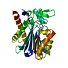
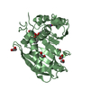
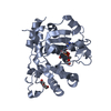
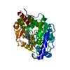
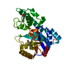
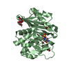
| Title | OXA-10 Avibactam complex with bound Iodide | ||||||
 Components Components | Beta-lactamase OXA-10 | ||||||
 Keywords Keywords | HYDROLASE / antibiotic resisitance | ||||||
| Function / homology |  Function and homology information Function and homology informationpenicillin binding / antibiotic catabolic process / cell wall organization / beta-lactamase activity / beta-lactamase / periplasmic space / response to antibiotic / plasma membrane Similarity search - Function | ||||||
| Biological species |   Pseudomonas aeruginosa (bacteria) Pseudomonas aeruginosa (bacteria) | ||||||
| Method |  X-RAY DIFFRACTION / SYNCHROTRON / MOLECULAR REPLACEMENT / molecular replacement / Resolution: 1.34 Å X-RAY DIFFRACTION / SYNCHROTRON / MOLECULAR REPLACEMENT / molecular replacement / Resolution: 1.34 Å | ||||||
 Authors Authors | Brem, J. | ||||||
 Citation Citation | Journal: Org. Biomol. Chem. / Year: 2017 Title: (13)C-Carbamylation as a mechanistic probe for the inhibition of class D beta-lactamases by avibactam and halide ions. Authors: Lohans, C.T. / Wang, D.Y. / Jorgensen, C. / Cahill, S.T. / Clifton, I.J. / McDonough, M.A. / Oswin, H.P. / Spencer, J. / Domene, C. / Claridge, T.D.W. / Brem, J. / Schofield, C.J. | ||||||
| History |
|
- Structure visualization
| Structure viewer | Molecule: MolmilJmol/JSmol |
|---|
- Downloads & links
-Download
| PDBx/mmCIF format | 5moz.cif.gz | 224.6 KB | Display | PDBx/mmCIF format |
|---|---|---|---|---|
| PDB format | pdb5moz.ent.gz | 177.1 KB | Display | PDB format |
| PDBx/mmJSON format | 5moz.json.gz | Tree view | PDBx/mmJSON format | |
| Others |  Other downloads Other downloads |
-Validation report
| Arichive directory | https://data.pdbj.org/pub/pdb/validation_reports/mo/5mozftp://data.pdbj.org/pub/pdb/validation_reports/mo/5moz | HTTPS FTP |
|---|
-Related structure data
| Related structure data | 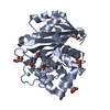 5mmyC 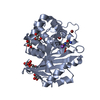 5mnuC 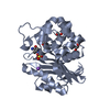 5moxC 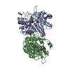 5fq9S S: Starting model for refinement C: citing same article ( |
|---|---|
| Similar structure data |


-Links
 PDBj
PDBj- Assembly
| Deposited unit | 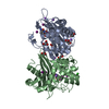
| ||||||||
|---|---|---|---|---|---|---|---|---|---|
| 1 | 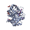
| ||||||||
| 2 | 
| ||||||||
| 3 |
| ||||||||
| Unit cell |
|
-Components
-Protein , 1 types, 2 molecules AB
| #1: Protein | Mass: 27655.488 Da / Num. of mol.: 2 Source method: isolated from a genetically manipulated source Source: (gene. exp.) Pseudomonas aeruginosa (bacteria) / Gene: bla, oxa10, pse2 / Production host: |
|---|
-Non-polymers , 6 types, 534 molecules 


| #2: Chemical |  Mass: 267.260 Da / Num. of mol.: 2 / Source method: obtained synthetically / Formula: C7H13N3O6S / Comment: antibiotic, inhibitor*YM Mass: 267.260 Da / Num. of mol.: 2 / Source method: obtained synthetically / Formula: C7H13N3O6S / Comment: antibiotic, inhibitor*YM#3: Chemical | ChemComp-GOL /  Mass: 92.094 Da / Num. of mol.: 6 / Source method: obtained synthetically / Formula: C3H8O3 Mass: 92.094 Da / Num. of mol.: 6 / Source method: obtained synthetically / Formula: C3H8O3#4: Chemical | ChemComp-NA /  Mass: 22.990 Da / Num. of mol.: 6 / Source method: obtained synthetically / Formula: Na Mass: 22.990 Da / Num. of mol.: 6 / Source method: obtained synthetically / Formula: Na#5: Chemical | ChemComp-IOD /  Mass: 126.904 Da / Num. of mol.: 8 / Source method: obtained synthetically / Formula: I Mass: 126.904 Da / Num. of mol.: 8 / Source method: obtained synthetically / Formula: I#6: Chemical | ChemComp-CL / |  Mass: 35.453 Da / Num. of mol.: 1 / Source method: obtained synthetically / Formula: Cl Mass: 35.453 Da / Num. of mol.: 1 / Source method: obtained synthetically / Formula: Cl#7: Water | ChemComp-HOH / | Mass: 18.015 Da / Num. of mol.: 511 / Source method: isolated from a natural source / Formula: H2O |
|---|
-Details
| Has protein modification | Y |
|---|
-Experimental details
-Experiment
| Experiment | Method: X-RAY DIFFRACTION / Number of used crystals: 1 |
|---|
- Sample preparation
| Crystal | Density Matthews: 3.2 Å3/Da / Density % sol: 61 % |
|---|---|
| Crystal grow | Temperature: 298 K / Method: vapor diffusion, sitting drop / pH: 6.5 / Details: 0.2M Sodium iodide, 20 % w/v PEG 3350 |
-Data collection
| Diffraction | Mean temperature: 100 K |
|---|---|
| Diffraction source | Source: SYNCHROTRON / Site: Diamond  / Beamline: I04 / Wavelength: 0.9795 Å / Beamline: I04 / Wavelength: 0.9795 Å |
| Detector | Type: DECTRIS PILATUS3 S 6M / Detector: PIXEL / Date: Jun 26, 2016 / Details: mirror |
| Radiation | Monochromator: double crystal / Protocol: SINGLE WAVELENGTH / Monochromatic (M) / Laue (L): M / Scattering type: x-ray |
| Radiation wavelength | Wavelength: 0.9795 Å / Relative weight: 1 |
| Reflection | Resolution: 1.34→79.29 Å / Num. obs: 141977 / % possible obs: 99.9 % / Observed criterion σ(I): 1.1 / Redundancy: 12.8 % / CC1/2: 0.999 / Rmerge(I) obs: 0.103 / Net I/σ(I): 11.2 |
| Reflection shell | Resolution: 1.34→1.36 Å / Redundancy: 12.1 % / Rmerge(I) obs: 2.828 / Mean I/σ(I) obs: 1.1 / Num. unique all: 6980 / CC1/2: 0.466 / % possible all: 99.2 |
-Phasing
| Phasing | Method: molecular replacement |
|---|
- Processing
| Software |
| ||||||||||||||||||
|---|---|---|---|---|---|---|---|---|---|---|---|---|---|---|---|---|---|---|---|
| Refinement | Method to determine structure: MOLECULAR REPLACEMENT Starting model: 5FQ9 Resolution: 1.34→79.29 Å / Cross valid method: FREE R-VALUE
| ||||||||||||||||||
| Displacement parameters | Biso max: 79.79 Å2 / Biso mean: 27.7582 Å2 / Biso min: 8.56 Å2 | ||||||||||||||||||
| Refinement step | Cycle: LAST / Resolution: 1.34→79.29 Å
|
