| Entry | Database: PDB / ID: 6gi1
|
|---|
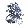
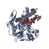
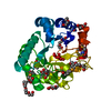
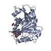
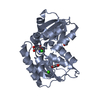
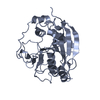
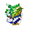
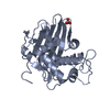
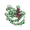
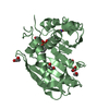
| Title | Crystal structure of the ferric enterobactin esterase (pfeE) mutant(S157A) from Pseudomonas aeruginosa in presence of enterobactin |
|---|
 Components Components | Ferric enterobactin esterase |
|---|
 Keywords Keywords | HYDROLASE / PfeE / PA2689 |
|---|
| Function / homology |  Function and homology information Function and homology information
iron(III)-enterobactin esterase / carboxylic ester hydrolase activity / hydrolase activity, acting on ester bonds / periplasmic spaceSimilarity search - Function |
|---|
| Biological species |  Pseudomonas aeruginosa PAO1 (bacteria) Pseudomonas aeruginosa PAO1 (bacteria) |
|---|
| Method |  X-RAY DIFFRACTION / SYNCHROTRON / MOLECULAR REPLACEMENT / Resolution: 1.66 Å X-RAY DIFFRACTION / SYNCHROTRON / MOLECULAR REPLACEMENT / Resolution: 1.66 Å |
|---|
 Authors Authors | Moynie, L. / Naismith, J.H. |
|---|
| Funding support |  United Kingdom, 1items United Kingdom, 1items | Organization | Grant number | Country |
|---|
| | United Kingdom |
|
|---|
 Citation Citation | Journal: ACS Chem. Biol. / Year: 2018
Title: A Key Role for the Periplasmic PfeE Esterase in Iron Acquisition via the Siderophore Enterobactin in Pseudomonas aeruginosa.
Authors: Perraud, Q. / Moynie, L. / Gasser, V. / Munier, M. / Godet, J. / Hoegy, F. / Mely, Y. / Mislin, G.L.A. / Naismith, J.H. / Schalk, I.J. |
|---|
| History | | Deposition | May 9, 2018 | Deposition site: PDBE / Processing site: PDBE |
|---|
| Revision 1.0 | Jun 20, 2018 | Provider: repository / Type: Initial release |
|---|
| Revision 1.1 | Aug 22, 2018 | Group: Data collection / Database references / Category: citation / citation_author
Item: _citation.country / _citation.journal_abbrev ..._citation.country / _citation.journal_abbrev / _citation.journal_id_CSD / _citation.journal_id_ISSN / _citation.pdbx_database_id_DOI / _citation.pdbx_database_id_PubMed / _citation.title / _citation.year |
|---|
| Revision 1.2 | Oct 3, 2018 | Group: Data collection / Database references / Category: citation / citation_author
Item: _citation.journal_volume / _citation.page_first ..._citation.journal_volume / _citation.page_first / _citation.page_last / _citation.title / _citation_author.identifier_ORCID |
|---|
| Revision 1.3 | May 15, 2024 | Group: Data collection / Database references / Refinement description
Category: chem_comp_atom / chem_comp_bond ...chem_comp_atom / chem_comp_bond / database_2 / struct_ncs_dom_lim
Item: _database_2.pdbx_DOI / _database_2.pdbx_database_accession ..._database_2.pdbx_DOI / _database_2.pdbx_database_accession / _struct_ncs_dom_lim.beg_auth_comp_id / _struct_ncs_dom_lim.beg_label_asym_id / _struct_ncs_dom_lim.beg_label_comp_id / _struct_ncs_dom_lim.beg_label_seq_id / _struct_ncs_dom_lim.end_auth_comp_id / _struct_ncs_dom_lim.end_label_asym_id / _struct_ncs_dom_lim.end_label_comp_id / _struct_ncs_dom_lim.end_label_seq_id |
|---|
|
|---|
 Movie
Movie Controller

 Yorodumi
 Open data
 Basic information
 Structure visualization
 Downloads & links
 Other downloads
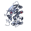


 PDBj
PDBj
 Assembly
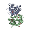
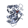


 Mass: 55.845 Da / Num. of mol.: 2 / Source method: obtained synthetically / Formula: Fe
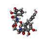
 Type: L-peptide linking / Mass: 241.197 Da / Num. of mol.: 1 / Source method: obtained synthetically / Formula: C10H11NO6
 Mass: 62.005 Da / Num. of mol.: 2 / Source method: obtained synthetically / Formula: NO3
 Mass: 62.068 Da / Num. of mol.: 5 / Source method: obtained synthetically / Formula: C2H6O2
 Mass: 669.546 Da / Num. of mol.: 1 / Source method: obtained synthetically / Formula: C30H27N3O15
 Sample preparation
 Processing