Mass: 18.015 Da / Num. of mol.: 266 / Source method: isolated from a natural source / Formula: H2O
-
Details
Has protein modification
Y
-
Experimental details
-
Experiment
Experiment
Method: X-RAY DIFFRACTION / Number of used crystals: 1
-
Sample preparation
Crystal
Density Matthews: 2.25 Å3/Da / Density % sol: 45.37 %
Crystal grow
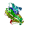
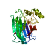
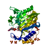
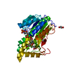
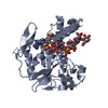
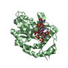
Temperature: 293 K / Method: microbatch / pH: 5.5 Details: Crystals of the 3-OST-1/PAP/heptasaccharide complex were obtained using micro-batch technique, by mixing 1.5 mL of the complex (12.9 mg/mL 3-OST-1, 5 mM heptasaccharide, 4 mM PAP, 23.6 mM ...Details: Crystals of the 3-OST-1/PAP/heptasaccharide complex were obtained using micro-batch technique, by mixing 1.5 mL of the complex (12.9 mg/mL 3-OST-1, 5 mM heptasaccharide, 4 mM PAP, 23.6 mM Tris pH 7.5, 142 mM NaCl) was mixed with 2.5 mL of 0.1 M sodium citrate pH 5.5 and 20% PEG 3000. Crystals grew to a usable size after 10 days incubation at room temperature. The crystals were transferred in two steps to a cryoprotectant solution containing 0.1 M sodium citrate pH 5.5, 0.1 M NaCl, 4 mM PAP, 20 mM heptasaccharide, 30% PEG3000, and 7.6% ethylene glycol. , temperature 293K
In the structure databanks used in Yorodumi, some data are registered as the other names, "COVID-19 virus" and "2019-nCoV". Here are the details of the virus and the list of structure data.
Jan 31, 2019. EMDB accession codes are about to change! (news from PDBe EMDB page)
EMDB accession codes are about to change! (news from PDBe EMDB page)
The allocation of 4 digits for EMDB accession codes will soon come to an end. Whilst these codes will remain in use, new EMDB accession codes will include an additional digit and will expand incrementally as the available range of codes is exhausted. The current 4-digit format prefixed with “EMD-” (i.e. EMD-XXXX) will advance to a 5-digit format (i.e. EMD-XXXXX), and so on. It is currently estimated that the 4-digit codes will be depleted around Spring 2019, at which point the 5-digit format will come into force.
The EM Navigator/Yorodumi systems omit the EMD- prefix.
Related info.:Q: What is EMD? / ID/Accession-code notation in Yorodumi/EM Navigator
Yorodumi is a browser for structure data from EMDB, PDB, SASBDB, etc.
This page is also the successor to EM Navigator detail page, and also detail information page/front-end page for Omokage search.
The word "yorodu" (or yorozu) is an old Japanese word meaning "ten thousand". "mi" (miru) is to see.
Related info.:EMDB / PDB / SASBDB / Comparison of 3 databanks / Yorodumi Search / Aug 31, 2016. New EM Navigator & Yorodumi / Yorodumi Papers / Jmol/JSmol / Function and homology information / Changes in new EM Navigator and Yorodumi
 Movie
Movie Controller

 Yorodumi
 Open data
 Basic information
 Components
 Keywords
 Function and homology information
 X-RAY DIFFRACTION /
  Authors
 Citation
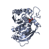
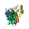
 Structure visualization
 Downloads & links
 Other downloads
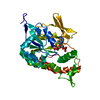


 PDBj
PDBj
 Assembly
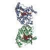

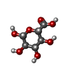
 Type: D-saccharide, beta linking / Mass: 194.139 Da / Num. of mol.: 1
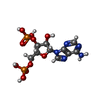

 Type: RNA linking / Mass: 427.201 Da / Num. of mol.: 2 / Source method: obtained synthetically / Formula: C10H15N5O10P2
 Sample preparation
 / Beamline: 22-ID / Wavelength: 1 Å
 Processing