 Movie
Movie Controller

[English] 日本語
 Yorodumi
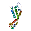
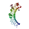
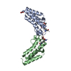
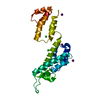
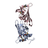
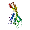
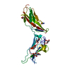
- PDB-5ewx: Fusion protein of T4 lysozyme and B4 domain of protein A from sta... -
+ Open data

- Basic information
| Entry | Database: PDB / ID: 5ewx | ||||||
|---|---|---|---|---|---|---|---|
| Title | Fusion protein of T4 lysozyme and B4 domain of protein A from staphylococcal aureus with chemical cross-linker EY-CBS | ||||||
 Components Components | Endolysin,Immunoglobulin G-binding protein A,Endolysin | ||||||
 Keywords Keywords | PROTEIN BINDING / Fusion / EY-CBS / alpha helix / cross-linker | ||||||
| Function / homology |  Function and homology information Function and homology informationIgG binding / viral release from host cell by cytolysis / peptidoglycan catabolic process / cell wall macromolecule catabolic process / lysozyme / lysozyme activity / host cell cytoplasm / defense response to bacterium Similarity search - Function | ||||||
| Biological species |  Enterobacteria phage T4 (virus) Enterobacteria phage T4 (virus)  Staphylococcus aureus (bacteria) Staphylococcus aureus (bacteria) | ||||||
| Method |  X-RAY DIFFRACTION / SYNCHROTRON / MOLECULAR REPLACEMENT / Resolution: 2.6 Å X-RAY DIFFRACTION / SYNCHROTRON / MOLECULAR REPLACEMENT / Resolution: 2.6 Å | ||||||
 Authors Authors | Jeong, W.H. / Lee, H. / Song, D.H. / Lee, J.O. | ||||||
 Citation Citation | Journal: Nat Commun / Year: 2016 Title: Connecting two proteins using a fusion alpha helix stabilized by a chemical cross linker. Authors: Jeong, W.H. / Lee, H. / Song, D.H. / Eom, J.H. / Kim, S.C. / Lee, H.S. / Lee, H. / Lee, J.O. | ||||||
| History |
|
- Structure visualization
| Structure viewer | Molecule: MolmilJmol/JSmol |
|---|
- Downloads & links
-Download
| PDBx/mmCIF format | 5ewx.cif.gz | 185.7 KB | Display | PDBx/mmCIF format |
|---|---|---|---|---|
| PDB format | pdb5ewx.ent.gz | 147.8 KB | Display | PDB format |
| PDBx/mmJSON format | 5ewx.json.gz | Tree view | PDBx/mmJSON format | |
| Others |  Other downloads Other downloads |
-Validation report
| Arichive directory | https://data.pdbj.org/pub/pdb/validation_reports/ew/5ewxftp://data.pdbj.org/pub/pdb/validation_reports/ew/5ewx | HTTPS FTP |
|---|
-Related structure data
| Related structure data |  5cbnC 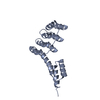 5cboC  5cocC  1deeS 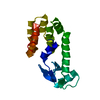 1lydS S: Starting model for refinement C: citing same article ( |
|---|---|
| Similar structure data |


-Links
 PDBj
PDBj


- Assembly
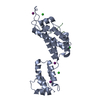
| Deposited unit | 
| ||||||||
|---|---|---|---|---|---|---|---|---|---|
| 1 | 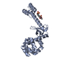
| ||||||||
| 2 | 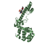
| ||||||||
| Unit cell |
|
-Components
| #1: Antibody | Mass: 25212.469 Da / Num. of mol.: 2 / Fragment: UNP RESIDUES 1-35, 219-266, 38-164 Mutation: A1212V, G1240A, E1258C, K1261A, L1262A, N40C, C54T, C97A, K162A Source method: isolated from a genetically manipulated source Details: The fusion protein of T4 lysozyme (residues 1-35), linker GGGGSGGGGS, protein A (B4 domain, residues 212-266), and T4 lysozyme (residues 38-164) Source: (gene. exp.) Enterobacteria phage T4 (virus), (gene. exp.) Staphylococcus aureus (bacteria)Gene: spa / Production host: #2: Chemical |   Mass: 521.348 Da / Num. of mol.: 2 / Source method: obtained synthetically / Formula: C18H14Cl2N2O8S2 Mass: 521.348 Da / Num. of mol.: 2 / Source method: obtained synthetically / Formula: C18H14Cl2N2O8S2#3: Water | ChemComp-HOH / |  Mass: 18.015 Da / Num. of mol.: 64 / Source method: isolated from a natural source / Formula: H2O Mass: 18.015 Da / Num. of mol.: 64 / Source method: isolated from a natural source / Formula: H2OSequence details | THE AUTHOR STATES THAT THERE ARE ERRORS AT THESE POSITIONS IN THE SEQUENCE DATABASE | |
|---|
-Experimental details
-Experiment
| Experiment | Method: X-RAY DIFFRACTION |
|---|
- Sample preparation
| Crystal | Density Matthews: 3.23 Å3/Da / Density % sol: 61.97 % Description: THE ENTRY CONTAINS FRIEDEL PAIRS IN I_PLUS/MINUS COLUMNS. square rod-plate |
|---|---|
| Crystal grow | Temperature: 277 K / Method: evaporation / pH: 7.5 / Details: 1.84M Na/K Phosphate, pH 7.5 |
-Data collection
| Diffraction | Mean temperature: 77 K |
|---|---|
| Diffraction source | Source: SYNCHROTRON / Site: PAL/PLS  / Beamline: 7A (6B, 6C1) / Wavelength: 0.9794 Å / Beamline: 7A (6B, 6C1) / Wavelength: 0.9794 Å |
| Detector | Type: ADSC QUANTUM 315 / Detector: CCD / Date: Oct 8, 2015 |
| Radiation | Protocol: SINGLE WAVELENGTH / Monochromatic (M) / Laue (L): M / Scattering type: x-ray |
| Radiation wavelength | Wavelength: 0.9794 Å / Relative weight: 1 |
| Reflection | Resolution: 2.6→50 Å / Num. obs: 35397 / % possible obs: 99.1 % / Redundancy: 3.5 % / Net I/σ(I): 18.3 |
- Processing
| Software |
| ||||||||||||||||||||||||||||||||||||||||||||||||||||||||||||||||||||||||||||||||||||||||||||||||||||||||||||||||||||||||||||||||||||||||||||||||||||||||||||||||||||||||||||||||||||||
|---|---|---|---|---|---|---|---|---|---|---|---|---|---|---|---|---|---|---|---|---|---|---|---|---|---|---|---|---|---|---|---|---|---|---|---|---|---|---|---|---|---|---|---|---|---|---|---|---|---|---|---|---|---|---|---|---|---|---|---|---|---|---|---|---|---|---|---|---|---|---|---|---|---|---|---|---|---|---|---|---|---|---|---|---|---|---|---|---|---|---|---|---|---|---|---|---|---|---|---|---|---|---|---|---|---|---|---|---|---|---|---|---|---|---|---|---|---|---|---|---|---|---|---|---|---|---|---|---|---|---|---|---|---|---|---|---|---|---|---|---|---|---|---|---|---|---|---|---|---|---|---|---|---|---|---|---|---|---|---|---|---|---|---|---|---|---|---|---|---|---|---|---|---|---|---|---|---|---|---|---|---|---|---|
| Refinement | Method to determine structure: MOLECULAR REPLACEMENT Starting model: 1LYD, 1DEE Resolution: 2.6→42.745 Å / SU ML: 0.39 / Cross valid method: FREE R-VALUE / σ(F): 1.39 / Phase error: 31.46 / Stereochemistry target values: ML
| ||||||||||||||||||||||||||||||||||||||||||||||||||||||||||||||||||||||||||||||||||||||||||||||||||||||||||||||||||||||||||||||||||||||||||||||||||||||||||||||||||||||||||||||||||||||
| Solvent computation | Shrinkage radii: 0.9 Å / VDW probe radii: 1.11 Å / Solvent model: FLAT BULK SOLVENT MODEL | ||||||||||||||||||||||||||||||||||||||||||||||||||||||||||||||||||||||||||||||||||||||||||||||||||||||||||||||||||||||||||||||||||||||||||||||||||||||||||||||||||||||||||||||||||||||
| Refinement step | Cycle: LAST / Resolution: 2.6→42.745 Å
| ||||||||||||||||||||||||||||||||||||||||||||||||||||||||||||||||||||||||||||||||||||||||||||||||||||||||||||||||||||||||||||||||||||||||||||||||||||||||||||||||||||||||||||||||||||||
| Refine LS restraints |
| ||||||||||||||||||||||||||||||||||||||||||||||||||||||||||||||||||||||||||||||||||||||||||||||||||||||||||||||||||||||||||||||||||||||||||||||||||||||||||||||||||||||||||||||||||||||
| LS refinement shell |
| ||||||||||||||||||||||||||||||||||||||||||||||||||||||||||||||||||||||||||||||||||||||||||||||||||||||||||||||||||||||||||||||||||||||||||||||||||||||||||||||||||||||||||||||||||||||
| Refinement TLS params. | Method: refined / Refine-ID: X-RAY DIFFRACTION
| ||||||||||||||||||||||||||||||||||||||||||||||||||||||||||||||||||||||||||||||||||||||||||||||||||||||||||||||||||||||||||||||||||||||||||||||||||||||||||||||||||||||||||||||||||||||
| Refinement TLS group |
|
