 Movie
Movie Controller

[English] 日本語
 Yorodumi
- PDB-5coc: Fusion protein of human calmodulin and B4 domain of protein A fro... -
+ Open data

- Basic information
| Entry | Database: PDB / ID: 5coc | ||||||
|---|---|---|---|---|---|---|---|
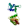
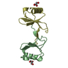
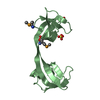
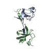
| Title | Fusion protein of human calmodulin and B4 domain of protein A from staphylococcal aureus | ||||||
 Components Components | Immunoglobulin G-binding protein A,Calmodulin | ||||||
 Keywords Keywords | PROTEIN BINDING / Fusion / alpha helix / cross-linker | ||||||
| Function / homology |  Function and homology information Function and homology information: / : / : / : / positive regulation of protein autophosphorylation / : / negative regulation of peptidyl-threonine phosphorylation / : / type 3 metabotropic glutamate receptor binding / IgG binding ...: / : / : / : / positive regulation of protein autophosphorylation / : / negative regulation of peptidyl-threonine phosphorylation / : / type 3 metabotropic glutamate receptor binding / IgG binding / positive regulation of DNA binding / positive regulation of peptidyl-threonine phosphorylation / negative regulation of high voltage-gated calcium channel activity / response to corticosterone / negative regulation of ryanodine-sensitive calcium-release channel activity / regulation of synaptic vesicle exocytosis / negative regulation of calcium ion export across plasma membrane / regulation of cardiac muscle cell action potential / positive regulation of protein serine/threonine kinase activity / nitric-oxide synthase binding / regulation of cell communication by electrical coupling involved in cardiac conduction / adenylate cyclase binding / protein phosphatase activator activity / regulation of synaptic vesicle endocytosis / detection of calcium ion / catalytic complex / regulation of cardiac muscle contraction / positive regulation of nitric-oxide synthase activity / phosphatidylinositol 3-kinase binding / activation of adenylate cyclase activity / regulation of release of sequestered calcium ion into cytosol by sarcoplasmic reticulum / enzyme regulator activity / regulation of calcium-mediated signaling / titin binding / regulation of cardiac muscle contraction by regulation of the release of sequestered calcium ion / voltage-gated potassium channel complex / calcium channel complex / substantia nigra development / regulation of heart rate / response to amphetamine / nitric-oxide synthase regulator activity / adenylate cyclase activator activity / protein serine/threonine kinase activator activity / regulation of cytokinesis / spindle microtubule / sarcomere / response to calcium ion / G2/M transition of mitotic cell cycle / mitochondrial membrane / spindle pole / disordered domain specific binding / calcium-dependent protein binding / myelin sheath / synaptic vesicle membrane / growth cone / vesicle / transmembrane transporter binding / G protein-coupled receptor signaling pathway / protein domain specific binding / calcium ion binding / centrosome / protein kinase binding / protein-containing complex / nucleus / plasma membrane / cytoplasm Similarity search - Function | ||||||
| Biological species |   Staphylococcus aureus (bacteria) Staphylococcus aureus (bacteria) Homo sapiens (human) Homo sapiens (human) | ||||||
| Method |  X-RAY DIFFRACTION / SYNCHROTRON / MOLECULAR REPLACEMENT / molecular replacement / Resolution: 2.6691 Å X-RAY DIFFRACTION / SYNCHROTRON / MOLECULAR REPLACEMENT / molecular replacement / Resolution: 2.6691 Å | ||||||
 Authors Authors | Jeong, W.H. / Lee, H. / Song, D.H. / Lee, J.O. | ||||||
 Citation Citation | Journal: Nat Commun / Year: 2016 Title: Connecting two proteins using a fusion alpha helix stabilized by a chemical cross linker. Authors: Jeong, W.H. / Lee, H. / Song, D.H. / Eom, J.H. / Kim, S.C. / Lee, H.S. / Lee, H. / Lee, J.O. | ||||||
| History |
|
- Structure visualization
| Structure viewer | Molecule: MolmilJmol/JSmol |
|---|
- Downloads & links
-Download
| PDBx/mmCIF format | 5coc.cif.gz | 63.7 KB | Display | PDBx/mmCIF format |
|---|---|---|---|---|
| PDB format | pdb5coc.ent.gz | 45.4 KB | Display | PDB format |
| PDBx/mmJSON format | 5coc.json.gz | Tree view | PDBx/mmJSON format | |
| Others |  Other downloads Other downloads |
-Validation report
| Arichive directory | https://data.pdbj.org/pub/pdb/validation_reports/co/5cocftp://data.pdbj.org/pub/pdb/validation_reports/co/5coc | HTTPS FTP |
|---|
-Related structure data
| Related structure data |  5cbnC  5cboC  5ewxC  1cllS  2spzS S: Starting model for refinement C: citing same article ( |
|---|---|
| Similar structure data |


-Links
 PDBj
PDBj


- Assembly
| Deposited unit | 
| ||||||||
|---|---|---|---|---|---|---|---|---|---|
| 1 |
| ||||||||
| Unit cell |
|
-Components
| #1: Antibody | Mass: 14550.117 Da / Num. of mol.: 1 Fragment: B4 domain (UNP RESIDUES 213-267),N-terminal (UNP RESIDUES 5-78) Mutation: G240A, K261C, L1005A, T1006A, Q1009C Source method: isolated from a genetically manipulated source Details: The fusion protein of residues 213-267 from Immunoglobulin G-binding protein A and residues 5-78 of Calmodulin Source: (gene. exp.) Staphylococcus aureus (bacteria), (gene. exp.) Homo sapiens (human)Gene: spa, CALM1, CALM, CAM, CAM1, CALM2, CAM2, CAMB, CALM3, CALML2, CAM3, CAMC, CAMIII Production host: | ||||
|---|---|---|---|---|---|
| #2: Chemical |   Mass: 40.078 Da / Num. of mol.: 2 / Source method: obtained synthetically / Formula: Ca Mass: 40.078 Da / Num. of mol.: 2 / Source method: obtained synthetically / Formula: Ca#3: Water | ChemComp-HOH / |  Mass: 18.015 Da / Num. of mol.: 12 / Source method: isolated from a natural source / Formula: H2O Mass: 18.015 Da / Num. of mol.: 12 / Source method: isolated from a natural source / Formula: H2OHas protein modification | Y | |
-Experimental details
-Experiment
| Experiment | Method: X-RAY DIFFRACTION / Number of used crystals: 1 |
|---|
- Sample preparation
| Crystal | Density Matthews: 2.8 Å3/Da / Density % sol: 56.04 % |
|---|---|
| Crystal grow | Temperature: 298 K / Method: evaporation / Details: 10% w/v PEG 1000, 10% w/v PEG 8000 |
-Data collection
| Diffraction | Mean temperature: 77 K | ||||||||||||||||||||||||||||||||||||||||||||||||||||||||||||||||||
|---|---|---|---|---|---|---|---|---|---|---|---|---|---|---|---|---|---|---|---|---|---|---|---|---|---|---|---|---|---|---|---|---|---|---|---|---|---|---|---|---|---|---|---|---|---|---|---|---|---|---|---|---|---|---|---|---|---|---|---|---|---|---|---|---|---|---|---|
| Diffraction source | Source: SYNCHROTRON / Site: PAL/PLS  / Beamline: 7A (6B, 6C1) / Wavelength: 0.97934 Å / Beamline: 7A (6B, 6C1) / Wavelength: 0.97934 Å | ||||||||||||||||||||||||||||||||||||||||||||||||||||||||||||||||||
| Detector | Type: ADSC QUANTUM 270 / Detector: CCD / Date: Jun 13, 2014 | ||||||||||||||||||||||||||||||||||||||||||||||||||||||||||||||||||
| Radiation | Protocol: SINGLE WAVELENGTH / Monochromatic (M) / Laue (L): M / Scattering type: x-ray | ||||||||||||||||||||||||||||||||||||||||||||||||||||||||||||||||||
| Radiation wavelength | Wavelength: 0.97934 Å / Relative weight: 1 | ||||||||||||||||||||||||||||||||||||||||||||||||||||||||||||||||||
| Reflection | Resolution: 2.6691→50 Å / Num. obs: 4463 / % possible obs: 99.6 % / Redundancy: 5.9 % / Rmerge(I) obs: 0.099 / Χ2: 1.641 / Net I/av σ(I): 22.74 / Net I/σ(I): 10.4 / Num. measured all: 26545 | ||||||||||||||||||||||||||||||||||||||||||||||||||||||||||||||||||
| Reflection shell | Diffraction-ID: 1 / Rejects: _
|
-Phasing
| Phasing | Method: molecular replacement |
|---|
- Processing
| Software |
| ||||||||||||||||||||||||||||||||||||||||
|---|---|---|---|---|---|---|---|---|---|---|---|---|---|---|---|---|---|---|---|---|---|---|---|---|---|---|---|---|---|---|---|---|---|---|---|---|---|---|---|---|---|
| Refinement | Method to determine structure: MOLECULAR REPLACEMENT Starting model: 2SPZ, 1CLL Resolution: 2.6691→31.246 Å / SU ML: 0.38 / Cross valid method: FREE R-VALUE / σ(F): 1.41 / Phase error: 27.71 / Stereochemistry target values: ML
| ||||||||||||||||||||||||||||||||||||||||
| Solvent computation | Shrinkage radii: 0.9 Å / VDW probe radii: 1.11 Å / Solvent model: FLAT BULK SOLVENT MODEL | ||||||||||||||||||||||||||||||||||||||||
| Displacement parameters | Biso max: 183.16 Å2 / Biso mean: 65.4115 Å2 / Biso min: 20 Å2 | ||||||||||||||||||||||||||||||||||||||||
| Refinement step | Cycle: final / Resolution: 2.6691→31.246 Å
| ||||||||||||||||||||||||||||||||||||||||
| Refine LS restraints |
| ||||||||||||||||||||||||||||||||||||||||
| LS refinement shell | Refine-ID: X-RAY DIFFRACTION / Total num. of bins used: 3
| ||||||||||||||||||||||||||||||||||||||||
| Refinement TLS params. | Method: refined / Origin x: -7.1043 Å / Origin y: -6.3182 Å / Origin z: 4.9416 Å
| ||||||||||||||||||||||||||||||||||||||||
| Refinement TLS group | Selection details: chain A |
