 Movie
Movie Controller


+ Open data

- Basic information
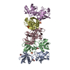
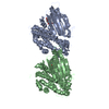
| Entry | Database: PDB / ID: 2x6l | |||||||||
|---|---|---|---|---|---|---|---|---|---|---|
| Title | X-ray Structure of Macrophage Inflammatory Protein-1 beta | |||||||||
 Components Components | C-C MOTIF CHEMOKINE 4 | |||||||||
 Keywords Keywords | IMMUNE SYSTEM / INFLAMMATORY RESPONSE / CHEMOTAXIS | |||||||||
| Function / homology |  Function and homology information Function and homology informationCCR1 chemokine receptor binding / positive regulation of natural killer cell chemotaxis / CCR5 chemokine receptor binding / CCR chemokine receptor binding / eosinophil chemotaxis / positive regulation of calcium ion transport / chemokine activity / Chemokine receptors bind chemokines / Interleukin-10 signaling / establishment or maintenance of cell polarity ...CCR1 chemokine receptor binding / positive regulation of natural killer cell chemotaxis / CCR5 chemokine receptor binding / CCR chemokine receptor binding / eosinophil chemotaxis / positive regulation of calcium ion transport / chemokine activity / Chemokine receptors bind chemokines / Interleukin-10 signaling / establishment or maintenance of cell polarity / host-mediated suppression of viral transcription / positive regulation of calcium-mediated signaling / cytokine activity / chemokine-mediated signaling pathway / response to toxic substance / response to virus / cell-cell signaling / antimicrobial humoral immune response mediated by antimicrobial peptide / G alpha (i) signalling events / cell adhesion / immune response / positive regulation of cell migration / inflammatory response / signal transduction / : / extracellular region / identical protein binding Similarity search - Function | |||||||||
| Biological species |  HOMO SAPIENS (human) HOMO SAPIENS (human) | |||||||||
| Method |  X-RAY DIFFRACTION / SYNCHROTRON / OTHER / Resolution: 2.602 Å X-RAY DIFFRACTION / SYNCHROTRON / OTHER / Resolution: 2.602 Å | |||||||||
 Authors Authors | Guo, Q. / Ren, M. / Tang, W. | |||||||||
 Citation Citation | Journal: Embo J. / Year: 2010 Title: Polymerization of Mip-1 Chemokine (Ccl3 and Ccl4) and Clearance of Mip-1 by Insulin-Degrading Enzyme. Authors: Ren, M. / Guo, Q. / Guo, L. / Lenz, M. / Qian, F. / Koenen, R.R. / Xu, H. / Schilling, A.B. / Weber, C. / Ye, R.D. / Dinner, A.R. / Tang, W. | |||||||||
| History |
|
- Structure visualization
| Structure viewer | Molecule: MolmilJmol/JSmol |
|---|
- Downloads & links
-Download
| PDBx/mmCIF format | 2x6l.cif.gz | 78.3 KB | Display | PDBx/mmCIF format |
|---|---|---|---|---|
| PDB format | pdb2x6l.ent.gz | 61 KB | Display | PDB format |
| PDBx/mmJSON format | 2x6l.json.gz | Tree view | PDBx/mmJSON format | |
| Others |  Other downloads Other downloads |
-Validation report
| Arichive directory | https://data.pdbj.org/pub/pdb/validation_reports/x6/2x6lftp://data.pdbj.org/pub/pdb/validation_reports/x6/2x6l | HTTPS FTP |
|---|
-Related structure data


-Links
 PDBj
PDBj

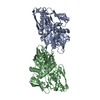
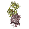
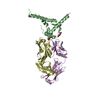
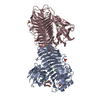
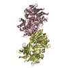
- Assembly
| Deposited unit | 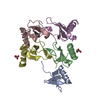
| ||||||||
|---|---|---|---|---|---|---|---|---|---|
| 1 | 
| ||||||||
| 2 | 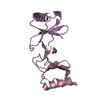
| ||||||||
| 3 | 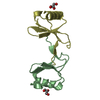
| ||||||||
| 4 | 
| ||||||||
| Unit cell |
| ||||||||
| Components on special symmetry positions |
|
-Components
| #1: Protein | Mass: 7824.742 Da / Num. of mol.: 5 / Source method: obtained synthetically / Source: (synth.) HOMO SAPIENS (human) / References: UniProt: P13236#2: Chemical |   Mass: 92.094 Da / Num. of mol.: 3 / Source method: obtained synthetically / Formula: C3H8O3 Mass: 92.094 Da / Num. of mol.: 3 / Source method: obtained synthetically / Formula: C3H8O3#3: Water | ChemComp-HOH / |  Mass: 18.015 Da / Num. of mol.: 145 / Source method: isolated from a natural source / Formula: H2O Mass: 18.015 Da / Num. of mol.: 145 / Source method: isolated from a natural source / Formula: H2OHas protein modification | Y | |
|---|
-Experimental details
-Experiment
| Experiment | Method: X-RAY DIFFRACTION |
|---|
- Sample preparation
| Crystal | Density Matthews: 3.11 Å3/Da / Density % sol: 60.2 % / Description: NONE |
|---|
-Data collection
| Diffraction | Mean temperature: 287 K |
|---|---|
| Diffraction source | Source: SYNCHROTRON / Site: APS  / Beamline: 19-ID / Wavelength: 0.9792 / Beamline: 19-ID / Wavelength: 0.9792 |
| Detector | Type: ADSC CCD / Detector: CCD / Date: Nov 1, 2001 |
| Radiation | Protocol: SINGLE WAVELENGTH / Monochromatic (M) / Laue (L): M / Scattering type: x-ray |
| Radiation wavelength | Wavelength: 0.9792 Å / Relative weight: 1 |
| Reflection | Resolution: 2.6→50 Å / Num. obs: 15529 / % possible obs: 100 % / Observed criterion σ(I): 1 / Redundancy: 7.2 % / Rmerge(I) obs: 0.12 / Net I/σ(I): 17.6 |
- Processing
| Software | Name: PHENIX / Version: (PHENIX.REFINE) / Classification: refinement | ||||||||||||||||||||||||||||||||||||||||||
|---|---|---|---|---|---|---|---|---|---|---|---|---|---|---|---|---|---|---|---|---|---|---|---|---|---|---|---|---|---|---|---|---|---|---|---|---|---|---|---|---|---|---|---|
| Refinement | Method to determine structure: OTHER Starting model: NONE Resolution: 2.602→49.448 Å / SU ML: 0.31 / σ(F): 0.16 / Phase error: 24.09 / Stereochemistry target values: ML
| ||||||||||||||||||||||||||||||||||||||||||
| Solvent computation | Shrinkage radii: 0.9 Å / VDW probe radii: 1.11 Å / Solvent model: FLAT BULK SOLVENT MODEL / Bsol: 42.241 Å2 / ksol: 0.405 e/Å3 | ||||||||||||||||||||||||||||||||||||||||||
| Displacement parameters |
| ||||||||||||||||||||||||||||||||||||||||||
| Refinement step | Cycle: LAST / Resolution: 2.602→49.448 Å
| ||||||||||||||||||||||||||||||||||||||||||
| Refine LS restraints |
| ||||||||||||||||||||||||||||||||||||||||||
| LS refinement shell |
|
