 Movie
Movie Controller

[English] 日本語
 Yorodumi
- PDB-5dib: 2.25 Angstrom resolution crystal structure of betaine aldehyde de... -
+ Open data

- Basic information
| Entry | Database: PDB / ID: 5dib | ||||||
|---|---|---|---|---|---|---|---|
| Title | 2.25 Angstrom resolution crystal structure of betaine aldehyde dehydrogenase (betB) Y450L point mutant from Staphylococcus aureus in complex with NAD+ and BME-modified Cys289 | ||||||
 Components Components | Betaine aldehyde dehydrogenase | ||||||
 Keywords Keywords | OXIDOREDUCTASE / BetB / structural genomics / NAD / NIAID / National Institute of Allergy and Infectious Diseases / CSGID / Rossmann fold / Center for Structural Genomics of Infectious Diseases | ||||||
| Function / homology |  Function and homology information Function and homology informationbetaine-aldehyde dehydrogenase (NAD+) activity / betaine-aldehyde dehydrogenase / glycine betaine biosynthetic process from choline / nucleotide binding / metal ion binding Similarity search - Function | ||||||
| Biological species |   Staphylococcus aureus (bacteria) Staphylococcus aureus (bacteria) | ||||||
| Method |  X-RAY DIFFRACTION / SYNCHROTRON / MOLECULAR REPLACEMENT / Resolution: 2.25 Å X-RAY DIFFRACTION / SYNCHROTRON / MOLECULAR REPLACEMENT / Resolution: 2.25 Å | ||||||
 Authors Authors | Halavaty, A.S. / Minasov, G. / Chen, C. / Joo, J.C. / Yakunin, A.F. / Anderson, W.F. / Center for Structural Genomics of Infectious Diseases (CSGID) | ||||||
 Citation Citation | Journal: To Be Published Title: 2.25 Angstrom resolution crystal structure of betaine aldehyde dehydrogenase (betB) Y450L point mutant from Staphylococcus aureus in complex with NAD+ and BME-modified Cys289 Authors: Halavaty, A.S. / Minasov, G. / Chen, C. / Joo, J.C. / Yakunin, A.F. / Anderson, W.F. / Center for Structural Genomics of Infectious Diseases (CSGID) #1: Journal: Appl. Environ. Microbiol. / Year: 2014Title: Structure-based mutational studies of substrate inhibition of betaine aldehyde dehydrogenase BetB from Staphylococcus aureus. Authors: Chen, C. / Joo, J.C. / Brown, G. / Stolnikova, E. / Halavaty, A.S. / Savchenko, A. / Anderson, W.F. / Yakunin, A.F. #2: Journal: Acta Crystallogr. D Biol. Crystallogr. / Year: 2015Title: Structural and functional analysis of betaine aldehyde dehydrogenase from Staphylococcus aureus. Authors: Halavaty, A.S. / Rich, R.L. / Chen, C. / Joo, J.C. / Minasov, G. / Dubrovska, I. / Winsor, J.R. / Myszka, D.G. / Duban, M. / Shuvalova, L. / Yakunin, A.F. / Anderson, W.F. | ||||||
| History |
|
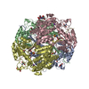
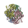
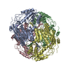
- Structure visualization
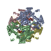
| Structure viewer | Molecule: MolmilJmol/JSmol |
|---|
- Downloads & links
-Download
| PDBx/mmCIF format | 5dib.cif.gz | 780.7 KB | Display | PDBx/mmCIF format |
|---|---|---|---|---|
| PDB format | pdb5dib.ent.gz | 648 KB | Display | PDB format |
| PDBx/mmJSON format | 5dib.json.gz | Tree view | PDBx/mmJSON format | |
| Others |  Other downloads Other downloads |
-Validation report
| Summary document | 5dib_validation.pdf.gz | 1.6 MB | Display | wwPDB validaton report |
|---|---|---|---|---|
| Full document | 5dib_full_validation.pdf.gz | 1.7 MB | Display | |
| Data in XML | 5dib_validation.xml.gz | 79.6 KB | Display | |
| Data in CIF | 5dib_validation.cif.gz | 110.6 KB | Display | |
| Arichive directory | https://data.pdbj.org/pub/pdb/validation_reports/di/5dibftp://data.pdbj.org/pub/pdb/validation_reports/di/5dib | HTTPS FTP |
-Related structure data
| Related structure data |  4mpbS S: Starting model for refinement |
|---|---|
| Similar structure data | |
| Other databases |


-Links
 PDBj
PDBj

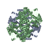
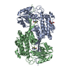
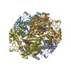
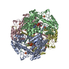
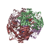
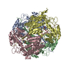
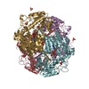
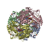
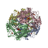
- Assembly
| Deposited unit | 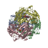
| ||||||||||||||||||||||||||||||||||||||||||||||||||||||||||||||||||||||||||||||||||||||||||||||||||||||||||||||||||||||||||||||||||||||||||||||||||||||
|---|---|---|---|---|---|---|---|---|---|---|---|---|---|---|---|---|---|---|---|---|---|---|---|---|---|---|---|---|---|---|---|---|---|---|---|---|---|---|---|---|---|---|---|---|---|---|---|---|---|---|---|---|---|---|---|---|---|---|---|---|---|---|---|---|---|---|---|---|---|---|---|---|---|---|---|---|---|---|---|---|---|---|---|---|---|---|---|---|---|---|---|---|---|---|---|---|---|---|---|---|---|---|---|---|---|---|---|---|---|---|---|---|---|---|---|---|---|---|---|---|---|---|---|---|---|---|---|---|---|---|---|---|---|---|---|---|---|---|---|---|---|---|---|---|---|---|---|---|---|---|---|
| 1 |
| ||||||||||||||||||||||||||||||||||||||||||||||||||||||||||||||||||||||||||||||||||||||||||||||||||||||||||||||||||||||||||||||||||||||||||||||||||||||
| Unit cell |
| ||||||||||||||||||||||||||||||||||||||||||||||||||||||||||||||||||||||||||||||||||||||||||||||||||||||||||||||||||||||||||||||||||||||||||||||||||||||
| Noncrystallographic symmetry (NCS) | NCS domain:
NCS domain segments: Component-ID: _ / Beg auth comp-ID: MET / Beg label comp-ID: MET / Refine code: _
NCS ensembles :
|
-Components
| #1: Protein | Mass: 57142.234 Da / Num. of mol.: 4 / Mutation: Y450L Source method: isolated from a genetically manipulated source Source: (gene. exp.) Staphylococcus aureus (bacteria)Gene: gbsA, CH51_13935, ERS157365_02065, ERS157366_01277, ERS157367_01590, ERS157368_01276, ERS157369_01327, ERS157370_01780, ERS157371_01447, ERS157372_01660, ERS157373_01815, ERS157374_00943, ...Gene: gbsA, CH51_13935, ERS157365_02065, ERS157366_01277, ERS157367_01590, ERS157368_01276, ERS157369_01327, ERS157370_01780, ERS157371_01447, ERS157372_01660, ERS157373_01815, ERS157374_00943, ERS157376_02019, ERS157379_02102, ERS157380_01277, ERS157381_01481, ERS157382_01327, ERS157383_01276, ERS157384_01683, ERS157385_01328, ERS157386_01590, ERS157387_01983, ERS157388_01278, ERS157389_01857, ERS157390_01571, ERS157391_01874, ERS157393_01280, ERS157394_01487, ERS157395_01059, ERS157409_01038, ERS195389_01428, ERS195391_02004, ERS445052_00785 Plasmid: P15TV-LIC / Production host: #2: Chemical | ChemComp-NAD /   Mass: 663.425 Da / Num. of mol.: 4 Mass: 663.425 Da / Num. of mol.: 4Source method: isolated from a genetically manipulated source Formula: C21H27N7O14P2 / Comment: NAD*YM #3: Chemical |   Mass: 22.990 Da / Num. of mol.: 3 / Source method: obtained synthetically / Formula: Na Mass: 22.990 Da / Num. of mol.: 3 / Source method: obtained synthetically / Formula: Na#4: Chemical |   Mass: 238.305 Da / Num. of mol.: 2 / Source method: obtained synthetically / Formula: C8H18N2O4S / Comment: pH buffer*YM Mass: 238.305 Da / Num. of mol.: 2 / Source method: obtained synthetically / Formula: C8H18N2O4S / Comment: pH buffer*YM#5: Water | ChemComp-HOH / |  Mass: 18.015 Da / Num. of mol.: 867 / Source method: isolated from a natural source / Formula: H2O Mass: 18.015 Da / Num. of mol.: 867 / Source method: isolated from a natural source / Formula: H2OHas protein modification | Y | |
|---|
-Experimental details
-Experiment
| Experiment | Method: X-RAY DIFFRACTION / Number of used crystals: 1 |
|---|
- Sample preparation
| Crystal | Density Matthews: 2.98 Å3/Da / Density % sol: 58.7 % |
|---|---|
| Crystal grow | Temperature: 295 K / Method: vapor diffusion, sitting drop / pH: 7 Details: Protein: BetB23 (Y450L mutant), 7 mg/ml in 10 mM Tris-HCl pH 8.3 500 mM NaCl 5 mM BME 5 mM NAD (co-crystallized) Crystallization: on 11/19/2014; The Classics II Suite (D3): 0.1 M HEPES pH 7. ...Details: Protein: BetB23 (Y450L mutant), 7 mg/ml in 10 mM Tris-HCl pH 8.3 500 mM NaCl 5 mM BME 5 mM NAD (co-crystallized) Crystallization: on 11/19/2014; The Classics II Suite (D3): 0.1 M HEPES pH 7.0 30% (v/v) Jeffamine ED-2001; soaked in crystallization conditions for cryo protection. |
-Data collection
| Diffraction | Mean temperature: 100 K |
|---|---|
| Diffraction source | Source: SYNCHROTRON / Site: APS  / Beamline: 21-ID-F / Wavelength: 0.97872 Å / Beamline: 21-ID-F / Wavelength: 0.97872 Å |
| Detector | Type: MARMOSAIC 225 mm CCD / Detector: CCD / Date: Jun 29, 2015 |
| Radiation | Monochromator: DIAMOND(111) / Protocol: SINGLE WAVELENGTH / Monochromatic (M) / Laue (L): M / Scattering type: x-ray |
| Radiation wavelength | Wavelength: 0.97872 Å / Relative weight: 1 |
| Reflection | Resolution: 2.25→29.72 Å / Num. obs: 122946 / % possible obs: 99.6 % / Redundancy: 3.7 % / Biso Wilson estimate: 30.3 Å2 / Rmerge(I) obs: 0.142 / Net I/σ(I): 9.4 |
| Reflection shell | Resolution: 2.25→2.29 Å / Redundancy: 3.6 % / Rmerge(I) obs: 0.649 / Mean I/σ(I) obs: 2.05 / % possible all: 99.8 |
- Processing
| Software |
| ||||||||||||||||||||||||||||||||||||||||||||||||||||||||||||||||||||||||||||||||||||||||||||||||||||||||||||||||||||||||||||||||||||||||||||||||||||||||||||||||||||||||||||||||||||||
|---|---|---|---|---|---|---|---|---|---|---|---|---|---|---|---|---|---|---|---|---|---|---|---|---|---|---|---|---|---|---|---|---|---|---|---|---|---|---|---|---|---|---|---|---|---|---|---|---|---|---|---|---|---|---|---|---|---|---|---|---|---|---|---|---|---|---|---|---|---|---|---|---|---|---|---|---|---|---|---|---|---|---|---|---|---|---|---|---|---|---|---|---|---|---|---|---|---|---|---|---|---|---|---|---|---|---|---|---|---|---|---|---|---|---|---|---|---|---|---|---|---|---|---|---|---|---|---|---|---|---|---|---|---|---|---|---|---|---|---|---|---|---|---|---|---|---|---|---|---|---|---|---|---|---|---|---|---|---|---|---|---|---|---|---|---|---|---|---|---|---|---|---|---|---|---|---|---|---|---|---|---|---|---|
| Refinement | Method to determine structure: MOLECULAR REPLACEMENT Starting model: 4mpb Resolution: 2.25→29.72 Å / Cor.coef. Fo:Fc: 0.925 / Cor.coef. Fo:Fc free: 0.892 / SU B: 14.051 / SU ML: 0.177 / Cross valid method: THROUGHOUT / ESU R: 0.284 / ESU R Free: 0.221 / Stereochemistry target values: MAXIMUM LIKELIHOOD / Details: HYDROGENS HAVE BEEN ADDED IN THE RIDING POSITIONS
| ||||||||||||||||||||||||||||||||||||||||||||||||||||||||||||||||||||||||||||||||||||||||||||||||||||||||||||||||||||||||||||||||||||||||||||||||||||||||||||||||||||||||||||||||||||||
| Solvent computation | Ion probe radii: 0.8 Å / Shrinkage radii: 0.8 Å / VDW probe radii: 1.2 Å / Solvent model: BABINET MODEL WITH MASK | ||||||||||||||||||||||||||||||||||||||||||||||||||||||||||||||||||||||||||||||||||||||||||||||||||||||||||||||||||||||||||||||||||||||||||||||||||||||||||||||||||||||||||||||||||||||
| Displacement parameters | Biso mean: 29.716 Å2
| ||||||||||||||||||||||||||||||||||||||||||||||||||||||||||||||||||||||||||||||||||||||||||||||||||||||||||||||||||||||||||||||||||||||||||||||||||||||||||||||||||||||||||||||||||||||
| Refinement step | Cycle: LAST / Resolution: 2.25→29.72 Å
| ||||||||||||||||||||||||||||||||||||||||||||||||||||||||||||||||||||||||||||||||||||||||||||||||||||||||||||||||||||||||||||||||||||||||||||||||||||||||||||||||||||||||||||||||||||||
| Refine LS restraints |
|