[English] 日本語
Yorodumi
- PDB-5gt6: Apo structure of Aldehyde Dehydrogenase from Bacillus cereus -

+
Open data


ID or keywords:

Loading...

-
Basic information

Entry
Database: PDB / ID: 5gt6
TitleApo structure of Aldehyde Dehydrogenase from Bacillus cereus
ComponentsBetaine-aldehyde dehydrogenase
KeywordsOXIDOREDUCTASE / Aldehyde Dehydrogenase NAD+ NADP+
Function / homology
Function and homology information


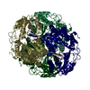
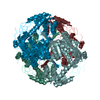
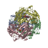
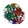
aldehyde dehydrogenase [NAD(P)+] / aldehyde dehydrogenase [NAD(P)+] activity / Oxidoreductases; Acting on the aldehyde or oxo group of donors; With NAD+ or NADP+ as acceptor
Similarity search - Function
Aldehyde Dehydrogenase; Chain A, domain 2 / Aldehyde Dehydrogenase; Chain A, domain 2 / Aldehyde Dehydrogenase; Chain A, domain 1 / Aldehyde Dehydrogenase; Chain A, domain 1 / Aldehyde dehydrogenase, glutamic acid active site / Aldehyde dehydrogenases glutamic acid active site. / Aldehyde dehydrogenase domain / Aldehyde dehydrogenase family / Aldehyde dehydrogenase, N-terminal / Aldehyde dehydrogenase, C-terminal ...Aldehyde Dehydrogenase; Chain A, domain 2 / Aldehyde Dehydrogenase; Chain A, domain 2 / Aldehyde Dehydrogenase; Chain A, domain 1 / Aldehyde Dehydrogenase; Chain A, domain 1 / Aldehyde dehydrogenase, glutamic acid active site / Aldehyde dehydrogenases glutamic acid active site. / Aldehyde dehydrogenase domain / Aldehyde dehydrogenase family / Aldehyde dehydrogenase, N-terminal / Aldehyde dehydrogenase, C-terminal / Aldehyde/histidinol dehydrogenase / 3-Layer(aba) Sandwich / Alpha Beta
Similarity search - Domain/homology
Aldehyde dehydrogenase (NAD) family protein
Similarity search - Component
Biological speciesBacillus cereus (bacteria)
MethodX-RAY DIFFRACTION / SYNCHROTRON / MOLECULAR REPLACEMENT / Resolution: 2.6 Å
AuthorsNgo, H.P.T. / Hong, S.H. / Ho, T.H. / Oh, D.K. / Kang, L.W.
CitationJournal: To Be Published
Title: crystal structures of aldehyde dehydrogenase from Bacillus cereus having atypical bidirectional oxidizing and reducing activities for all-trans-retinal
Authors: Ngo, H.P.T. / Hong, S.H. / Ho, T.H. / Oh, D.K. / Kang, L.W.
History
DepositionAug 18, 2016Deposition site: PDBJ / Processing site: PDBJ
SupersessionSep 6, 2017ID: 4PS9
Revision 1.0Sep 6, 2017Provider: repository / Type: Initial release
Revision 1.1Nov 8, 2023Group: Data collection / Database references ...Data collection / Database references / Derived calculations / Refinement description
Category: chem_comp_atom / chem_comp_bond ...chem_comp_atom / chem_comp_bond / database_2 / pdbx_initial_refinement_model / struct_conn
Item: _database_2.pdbx_DOI / _database_2.pdbx_database_accession ..._database_2.pdbx_DOI / _database_2.pdbx_database_accession / _struct_conn.pdbx_dist_value / _struct_conn.ptnr1_auth_asym_id / _struct_conn.ptnr1_auth_comp_id / _struct_conn.ptnr1_auth_seq_id / _struct_conn.ptnr1_label_asym_id / _struct_conn.ptnr1_label_atom_id / _struct_conn.ptnr1_label_comp_id / _struct_conn.ptnr1_label_seq_id / _struct_conn.ptnr2_auth_asym_id / _struct_conn.ptnr2_auth_comp_id / _struct_conn.ptnr2_auth_seq_id / _struct_conn.ptnr2_label_asym_id / _struct_conn.ptnr2_label_atom_id / _struct_conn.ptnr2_label_comp_id

-
Structure visualization

Structure viewerMolecule:
MolmilJmol/JSmol

Downloads & links

-
Assembly

Deposited unit
A: Betaine-aldehyde dehydrogenase
B: Betaine-aldehyde dehydrogenase
C: Betaine-aldehyde dehydrogenase
D: Betaine-aldehyde dehydrogenase
hetero molecules


Theoretical massNumber of molelcules
Total (without water)223,4328
Polymers223,3404
Non-polymers924
Water1,65792
1


  • Idetical with deposited unit
  • defined by author&software
TypeNameSymmetry operationNumber
identity operation1_555x,y,z1
Buried area18760 Å2
ΔGint-131 kcal/mol
Surface area61600 Å2
MethodPISA
Unit cell
Length a, b, c (Å)83.527, 93.284, 145.472
Angle α, β, γ (deg.)90.00, 98.05, 90.00
Int Tables number4
Space group name H-MP1211

-
Components

#1: Protein
Betaine-aldehyde dehydrogenase /


Mass: 55834.969 Da / Num. of mol.: 4
Source method: isolated from a genetically manipulated source
Source: (gene. exp.) Bacillus cereus (bacteria) / Gene: AT268_22120 / Production host: Escherichia coli (E. coli) / References: UniProt: A0A150BLG9
#2: Chemical
ChemComp-NA / SODIUM ION


Mass: 22.990 Da / Num. of mol.: 4 / Source method: obtained synthetically / Formula: Na
#3: Water ChemComp-HOH / water / Water


Mass: 18.015 Da / Num. of mol.: 92 / Source method: isolated from a natural source / Formula: H2O

-
Experimental details

-
Experiment

ExperimentMethod: X-RAY DIFFRACTION / Number of used crystals: 1

-
Sample preparation

CrystalDensity Matthews: 2.59 Å3/Da / Density % sol: 52.47 %
Crystal growTemperature: 287 K / Method: vapor diffusion, sitting drop / pH: 7 / Details: 0.15 M DL-malic acid pH 7.0, 18% PEG 3350

-
Data collection

DiffractionMean temperature: 100 K
Diffraction sourceSource: SYNCHROTRON / Site: PAL/PLS / Beamline: 5C (4A) / Wavelength: 0.97951 Å
DetectorType: ADSC QUANTUM 315r / Detector: CCD / Date: Nov 26, 2012
RadiationMonochromator: DCM Si (111) Crystal / Protocol: SINGLE WAVELENGTH / Monochromatic (M) / Laue (L): M / Scattering type: x-ray
Radiation wavelengthWavelength: 0.97951 Å / Relative weight: 1
ReflectionResolution: 2.6→50 Å / Num. obs: 67464 / % possible obs: 99.2 % / Observed criterion σ(I): 1 / Redundancy: 7.4 % / Rmerge(I) obs: 0.046 / Net I/σ(I): 69.7
Reflection shellResolution: 2.6→2.64 Å / Redundancy: 7.1 % / Rmerge(I) obs: 0.075 / % possible all: 97.7

-
Processing

Software
NameVersionClassification
REFMAC5.8.0135refinement
HKL-2000data reduction
HKL-2000data scaling
PHASERphasing
RefinementMethod to determine structure: MOLECULAR REPLACEMENT
Starting model: 4FR8
Resolution: 2.6→49.57 Å / Cor.coef. Fo:Fc: 0.93 / Cor.coef. Fo:Fc free: 0.867 / SU B: 9.881 / SU ML: 0.211 / Cross valid method: THROUGHOUT / ESU R: 1.059 / ESU R Free: 0.325 / Stereochemistry target values: MAXIMUM LIKELIHOOD / Details: HYDROGENS HAVE BEEN ADDED IN THE RIDING POSITIONS
RfactorNum. reflection% reflectionSelection details
Rfree0.25095 3423 5.1 %RANDOM
Rwork0.18201 ---
obs0.18548 64021 99.09 %-
Solvent computationIon probe radii: 0.8 Å / Shrinkage radii: 0.8 Å / VDW probe radii: 1.2 Å / Solvent model: MASK
Displacement parametersBiso mean: 21.36 Å2
Baniso -1Baniso -2Baniso -3
1--0.01 Å20 Å20.01 Å2
2--0 Å20 Å2
3---0.01 Å2
Refinement stepCycle: 1 / Resolution: 2.6→49.57 Å
ProteinNucleic acidLigandSolventTotal
Num. atoms15117 0 4 92 15213
Refine LS restraints
Refine-IDTypeDev idealDev ideal targetNumber
X-RAY DIFFRACTIONr_bond_refined_d0.0150.01915457
X-RAY DIFFRACTIONr_bond_other_d0.0020.0214695
X-RAY DIFFRACTIONr_angle_refined_deg1.781.95120943
X-RAY DIFFRACTIONr_angle_other_deg1.096333850
X-RAY DIFFRACTIONr_dihedral_angle_1_deg7.09851954
X-RAY DIFFRACTIONr_dihedral_angle_2_deg39.02325.472689
X-RAY DIFFRACTIONr_dihedral_angle_3_deg20.507152596
X-RAY DIFFRACTIONr_dihedral_angle_4_deg20.6281548
X-RAY DIFFRACTIONr_chiral_restr0.1150.22339
X-RAY DIFFRACTIONr_gen_planes_refined0.0070.02117674
X-RAY DIFFRACTIONr_gen_planes_other0.0020.023394
X-RAY DIFFRACTIONr_nbd_refined
X-RAY DIFFRACTIONr_nbd_other
X-RAY DIFFRACTIONr_nbtor_refined
X-RAY DIFFRACTIONr_nbtor_other
X-RAY DIFFRACTIONr_xyhbond_nbd_refined
X-RAY DIFFRACTIONr_xyhbond_nbd_other
X-RAY DIFFRACTIONr_metal_ion_refined
X-RAY DIFFRACTIONr_metal_ion_other
X-RAY DIFFRACTIONr_symmetry_vdw_refined
X-RAY DIFFRACTIONr_symmetry_vdw_other
X-RAY DIFFRACTIONr_symmetry_hbond_refined
X-RAY DIFFRACTIONr_symmetry_hbond_other
X-RAY DIFFRACTIONr_symmetry_metal_ion_refined
X-RAY DIFFRACTIONr_symmetry_metal_ion_other
X-RAY DIFFRACTIONr_mcbond_it1.2692.0167828
X-RAY DIFFRACTIONr_mcbond_other1.2682.0167827
X-RAY DIFFRACTIONr_mcangle_it2.0763.0219778
X-RAY DIFFRACTIONr_mcangle_other2.0773.0219779
X-RAY DIFFRACTIONr_scbond_it1.8252.2367629
X-RAY DIFFRACTIONr_scbond_other1.8252.2367630
X-RAY DIFFRACTIONr_scangle_it
X-RAY DIFFRACTIONr_scangle_other3.0533.25211166
X-RAY DIFFRACTIONr_long_range_B_refined4.04915.78316749
X-RAY DIFFRACTIONr_long_range_B_other4.0515.78616748
X-RAY DIFFRACTIONr_rigid_bond_restr
X-RAY DIFFRACTIONr_sphericity_free
X-RAY DIFFRACTIONr_sphericity_bonded
LS refinement shellResolution: 2.602→2.67 Å / Total num. of bins used: 20
RfactorNum. reflection% reflection
Rfree0.347 248 -
Rwork0.217 4576 -
obs--97.08 %

+
About Yorodumi

-
News

-
Feb 9, 2022. New format data for meta-information of EMDB entries

New format data for meta-information of EMDB entries

  • Version 3 of the EMDB header file is now the official format.
  • The previous official version 1.9 will be removed from the archive.

Related info.:EMDB header

External links:wwPDB to switch to version 3 of the EMDB data model

-
Aug 12, 2020. Covid-19 info

Covid-19 info

URL: https://pdbj.org/emnavi/covid19.php

New page: Covid-19 featured information page in EM Navigator.

Related info.:Covid-19 info / Mar 5, 2020. Novel coronavirus structure data

+
Mar 5, 2020. Novel coronavirus structure data

Novel coronavirus structure data

Related info.:Yorodumi Speices / Aug 12, 2020. Covid-19 info

External links:COVID-19 featured content - PDBj / Molecule of the Month (242):Coronavirus Proteases

+
Jan 31, 2019. EMDB accession codes are about to change! (news from PDBe EMDB page)

EMDB accession codes are about to change! (news from PDBe EMDB page)

  • The allocation of 4 digits for EMDB accession codes will soon come to an end. Whilst these codes will remain in use, new EMDB accession codes will include an additional digit and will expand incrementally as the available range of codes is exhausted. The current 4-digit format prefixed with “EMD-” (i.e. EMD-XXXX) will advance to a 5-digit format (i.e. EMD-XXXXX), and so on. It is currently estimated that the 4-digit codes will be depleted around Spring 2019, at which point the 5-digit format will come into force.
  • The EM Navigator/Yorodumi systems omit the EMD- prefix.

Related info.:Q: What is EMD? / ID/Accession-code notation in Yorodumi/EM Navigator

External links:EMDB Accession Codes are Changing Soon! / Contact to PDBj

+
Jul 12, 2017. Major update of PDB

Major update of PDB

  • wwPDB released updated PDB data conforming to the new PDBx/mmCIF dictionary.
  • This is a major update changing the version number from 4 to 5, and with Remediation, in which all the entries are updated.
  • In this update, many items about electron microscopy experimental information are reorganized (e.g. em_software).
  • Now, EM Navigator and Yorodumi are based on the updated data.

External links:wwPDB Remediation / Enriched Model Files Conforming to OneDep Data Standards Now Available in the PDB FTP Archive

-
Yorodumi

Thousand views of thousand structures

  • Yorodumi is a browser for structure data from EMDB, PDB, SASBDB, etc.
  • This page is also the successor to EM Navigator detail page, and also detail information page/front-end page for Omokage search.
  • The word "yorodu" (or yorozu) is an old Japanese word meaning "ten thousand". "mi" (miru) is to see.

Related info.:EMDB / PDB / SASBDB / Comparison of 3 databanks / Yorodumi Search / Aug 31, 2016. New EM Navigator & Yorodumi / Yorodumi Papers / Jmol/JSmol / Function and homology information / Changes in new EM Navigator and Yorodumi

Read more