 Movie
Movie Controller

[English] 日本語
 Yorodumi
- PDB-2o2p: Crystal structure of the C-terminal domain of rat 10'formyltetrah... -
+ Open data

- Basic information
| Entry | Database: PDB / ID: 2o2p | ||||||
|---|---|---|---|---|---|---|---|
| Title | Crystal structure of the C-terminal domain of rat 10'formyltetrahydrofolate dehydrogenase | ||||||
 Components Components | Formyltetrahydrofolate dehydrogenase | ||||||
 Keywords Keywords | OXIDOREDUCTASE / ALDEHYDE DEHYDROGENASE / FDH | ||||||
| Function / homology |  Function and homology information Function and homology informationMetabolism of folate and pterines / aldehyde dehydrogenase (NADP+) activity / formyltetrahydrofolate dehydrogenase / formyltetrahydrofolate dehydrogenase activity / 10-formyltetrahydrofolate catabolic process / aldehyde dehydrogenase [NAD(P)+] activity / NADPH regeneration / aldehyde dehydrogenase (NAD+) activity / tetrahydrofolate interconversion / folic acid metabolic process ...Metabolism of folate and pterines / aldehyde dehydrogenase (NADP+) activity / formyltetrahydrofolate dehydrogenase / formyltetrahydrofolate dehydrogenase activity / 10-formyltetrahydrofolate catabolic process / aldehyde dehydrogenase [NAD(P)+] activity / NADPH regeneration / aldehyde dehydrogenase (NAD+) activity / tetrahydrofolate interconversion / folic acid metabolic process / tetrahydrofolate biosynthetic process / protein-containing complex binding / protein-containing complex / cytosol Similarity search - Function | ||||||
| Biological species |  | ||||||
| Method |  X-RAY DIFFRACTION / SYNCHROTRON / FOURIER SYNTHESIS / Resolution: 1.7 Å X-RAY DIFFRACTION / SYNCHROTRON / FOURIER SYNTHESIS / Resolution: 1.7 Å | ||||||
 Authors Authors | Tsybovsky, Y. / Donato, H. / Krupenko, N.I. / Davies, C. / Krupenko, S.A. | ||||||
 Citation Citation | Journal: Biochemistry / Year: 2007 Title: Crystal structures of the carboxyl terminal domain of rat 10-formyltetrahydrofolate dehydrogenase: implications for the catalytic mechanism of aldehyde dehydrogenases. Authors: Tsybovsky, Y. / Donato, H. / Krupenko, N.I. / Davies, C. / Krupenko, S.A. | ||||||
| History |
|
- Structure visualization
| Structure viewer | Molecule: MolmilJmol/JSmol |
|---|
- Downloads & links
-Download
| PDBx/mmCIF format | 2o2p.cif.gz | 436.7 KB | Display | PDBx/mmCIF format |
|---|---|---|---|---|
| PDB format | pdb2o2p.ent.gz | 354.8 KB | Display | PDB format |
| PDBx/mmJSON format | 2o2p.json.gz | Tree view | PDBx/mmJSON format | |
| Others |  Other downloads Other downloads |
-Validation report
| Arichive directory | https://data.pdbj.org/pub/pdb/validation_reports/o2/2o2pftp://data.pdbj.org/pub/pdb/validation_reports/o2/2o2p | HTTPS FTP |
|---|
-Related structure data
| Related structure data |  2o2qSC  2o2rC S: Starting model for refinement C: citing same article ( |
|---|---|
| Similar structure data |


-Links
 PDBj
PDBj


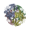
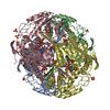
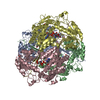
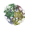
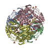
- Assembly
| Deposited unit | 
| ||||||||
|---|---|---|---|---|---|---|---|---|---|
| 1 |
| ||||||||
| 2 | 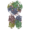
| ||||||||
| Unit cell |
| ||||||||
| Details | The biological unit is the tetramer |
-Components
| #1: Protein | Mass: 56621.547 Da / Num. of mol.: 4 / Fragment: C-terminal domain, residues 397-902 Source method: isolated from a genetically manipulated source Source: (gene. exp.)  References: UniProt: Q5HZB2, UniProt: P28037*PLUS, formyltetrahydrofolate dehydrogenase #2: Chemical | ChemComp-SO4 /   Mass: 96.063 Da / Num. of mol.: 28 / Source method: obtained synthetically / Formula: SO4 Mass: 96.063 Da / Num. of mol.: 28 / Source method: obtained synthetically / Formula: SO4#3: Chemical | ChemComp-GOL /   Mass: 92.094 Da / Num. of mol.: 4 / Source method: obtained synthetically / Formula: C3H8O3 Mass: 92.094 Da / Num. of mol.: 4 / Source method: obtained synthetically / Formula: C3H8O3#4: Water | ChemComp-HOH / |  Mass: 18.015 Da / Num. of mol.: 1795 / Source method: isolated from a natural source / Formula: H2O Mass: 18.015 Da / Num. of mol.: 1795 / Source method: isolated from a natural source / Formula: H2O |
|---|
-Experimental details
-Experiment
| Experiment | Method: X-RAY DIFFRACTION / Number of used crystals: 1 |
|---|
- Sample preparation
| Crystal grow | Temperature: 288 K / Method: vapor diffusion, hanging drop / pH: 7.5 Details: 1.4 M AMMONIUM SULPHATE, 0.1M TRIS, pH 7.5, VAPOR DIFFUSION, HANGING DROP, temperature 288K |
|---|
-Data collection
| Diffraction | Mean temperature: 100 K |
|---|---|
| Diffraction source | Source: SYNCHROTRON / Site: APS  / Beamline: 22-ID / Wavelength: 1 Å / Beamline: 22-ID / Wavelength: 1 Å |
| Detector | Type: MARMOSAIC 300 mm CCD / Detector: CCD / Date: Oct 22, 2005 |
| Radiation | Protocol: SINGLE WAVELENGTH / Monochromatic (M) / Laue (L): M / Scattering type: x-ray |
| Radiation wavelength | Wavelength: 1 Å / Relative weight: 1 |
| Reflection | Resolution: 1.7→50 Å / Num. all: 485421 / Num. obs: 485421 / % possible obs: 97.5 % / Observed criterion σ(F): 0 / Observed criterion σ(I): 0 / Redundancy: 5.1 % / Biso Wilson estimate: 19.4 Å2 / Rmerge(I) obs: 0.085 / Rsym value: 0.085 / Net I/σ(I): 10.1 |
| Reflection shell | Resolution: 1.7→1.76 Å / Redundancy: 4.8 % / Rmerge(I) obs: 0.558 / Mean I/σ(I) obs: 2.5 / Num. unique all: 47647 / Rsym value: 0.558 / % possible all: 95.8 |
- Processing
| Software |
| ||||||||||||||||||||||||||||||||||||||||||||||||||||||||||||||||||||||||||||||||||||||||||
|---|---|---|---|---|---|---|---|---|---|---|---|---|---|---|---|---|---|---|---|---|---|---|---|---|---|---|---|---|---|---|---|---|---|---|---|---|---|---|---|---|---|---|---|---|---|---|---|---|---|---|---|---|---|---|---|---|---|---|---|---|---|---|---|---|---|---|---|---|---|---|---|---|---|---|---|---|---|---|---|---|---|---|---|---|---|---|---|---|---|---|---|
| Refinement | Method to determine structure: FOURIER SYNTHESIS Starting model: PDB ENTRY 2O2Q Resolution: 1.7→50 Å / Cor.coef. Fo:Fc: 0.963 / Cor.coef. Fo:Fc free: 0.957 / SU B: 1.236 / SU ML: 0.041 / Isotropic thermal model: Isotropic / Cross valid method: THROUGHOUT / σ(F): 0 / σ(I): 0 / ESU R: 0.064 / ESU R Free: 0.064 / Stereochemistry target values: MAXIMUM LIKELIHOOD
| ||||||||||||||||||||||||||||||||||||||||||||||||||||||||||||||||||||||||||||||||||||||||||
| Solvent computation | Ion probe radii: 0.8 Å / Shrinkage radii: 0.8 Å / VDW probe radii: 1.4 Å / Solvent model: MASK | ||||||||||||||||||||||||||||||||||||||||||||||||||||||||||||||||||||||||||||||||||||||||||
| Displacement parameters | Biso mean: 19.203 Å2
| ||||||||||||||||||||||||||||||||||||||||||||||||||||||||||||||||||||||||||||||||||||||||||
| Refinement step | Cycle: LAST / Resolution: 1.7→50 Å
| ||||||||||||||||||||||||||||||||||||||||||||||||||||||||||||||||||||||||||||||||||||||||||
| Refine LS restraints |
| ||||||||||||||||||||||||||||||||||||||||||||||||||||||||||||||||||||||||||||||||||||||||||
| LS refinement shell | Resolution: 1.7→1.744 Å / Total num. of bins used: 20
|
