 Movie
Movie Controller

[English] 日本語
 Yorodumi
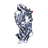
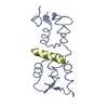
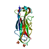
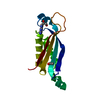
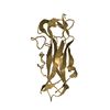
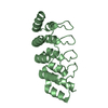
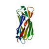
- PDB-5csd: Ligand binding domain 2 of Penicillium marneffei MP1 protein in c... -
+ Open data

- Basic information
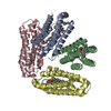
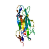
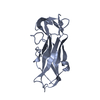
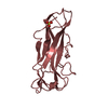
| Entry | Database: PDB / ID: 5csd | ||||||
|---|---|---|---|---|---|---|---|
| Title | Ligand binding domain 2 of Penicillium marneffei MP1 protein in complex with arachidonic acids | ||||||
 Components Components | Envelope glycoprotein | ||||||
 Keywords Keywords | LIPID BINDING PROTEIN / arachidonic acid / Ligand binding domain | ||||||
| Function / homology | Cell wall mannoprotein 1 / Hydrophobic surface binding protein A / fungal-type cell wall / lipid binding / extracellular region / ARACHIDONIC ACID / Cell wall mannoprotein 1 Function and homology information Function and homology information | ||||||
| Biological species |  Talaromyces marneffei PM1 (fungus) Talaromyces marneffei PM1 (fungus) | ||||||
| Method |  X-RAY DIFFRACTION / SYNCHROTRON / MOLECULAR REPLACEMENT / Resolution: 1.45 Å X-RAY DIFFRACTION / SYNCHROTRON / MOLECULAR REPLACEMENT / Resolution: 1.45 Å | ||||||
 Authors Authors | Lam, W.H. / Zhang, H. / Hao, Q. | ||||||
 Citation Citation | Journal: Cell Chem Biol / Year: 2017 Title: Talaromyces marneffei Mp1p Is a Virulence Factor that Binds and Sequesters a Key Proinflammatory Lipid to Dampen Host Innate Immune Response Authors: Sze, K.H. / Lam, W.H. / Zhang, H. / Ke, Y.H. / Tse, M.K. / Woo, P.C. / Lau, S.K. / Lau, C.C. / Cai, J.P. / Tung, E.T. / Lo, R.K. / Xu, S. / Kao, R.Y. / Hao, Q. / Yuen, K.Y. | ||||||
| History |
|
- Structure visualization
| Structure viewer | Molecule: MolmilJmol/JSmol |
|---|
- Downloads & links
-Download
| PDBx/mmCIF format | 5csd.cif.gz | 259.5 KB | Display | PDBx/mmCIF format |
|---|---|---|---|---|
| PDB format | pdb5csd.ent.gz | 210.8 KB | Display | PDB format |
| PDBx/mmJSON format | 5csd.json.gz | Tree view | PDBx/mmJSON format | |
| Others |  Other downloads Other downloads |
-Validation report
| Arichive directory | https://data.pdbj.org/pub/pdb/validation_reports/cs/5csdftp://data.pdbj.org/pub/pdb/validation_reports/cs/5csd | HTTPS FTP |
|---|
-Related structure data
-Links
 PDBj
PDBj
- Assembly
| Deposited unit | 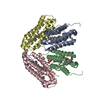
| ||||||||||||||||||||||||||||||||||||||||||||||||||||||||||||||||||||||||||||||||||||||||||||||||||||||||||||||||||||||||||||||||||||||||||||||||||||||
|---|---|---|---|---|---|---|---|---|---|---|---|---|---|---|---|---|---|---|---|---|---|---|---|---|---|---|---|---|---|---|---|---|---|---|---|---|---|---|---|---|---|---|---|---|---|---|---|---|---|---|---|---|---|---|---|---|---|---|---|---|---|---|---|---|---|---|---|---|---|---|---|---|---|---|---|---|---|---|---|---|---|---|---|---|---|---|---|---|---|---|---|---|---|---|---|---|---|---|---|---|---|---|---|---|---|---|---|---|---|---|---|---|---|---|---|---|---|---|---|---|---|---|---|---|---|---|---|---|---|---|---|---|---|---|---|---|---|---|---|---|---|---|---|---|---|---|---|---|---|---|---|
| 1 | 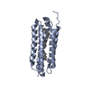
| ||||||||||||||||||||||||||||||||||||||||||||||||||||||||||||||||||||||||||||||||||||||||||||||||||||||||||||||||||||||||||||||||||||||||||||||||||||||
| 2 | 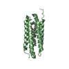
| ||||||||||||||||||||||||||||||||||||||||||||||||||||||||||||||||||||||||||||||||||||||||||||||||||||||||||||||||||||||||||||||||||||||||||||||||||||||
| 3 | 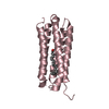
| ||||||||||||||||||||||||||||||||||||||||||||||||||||||||||||||||||||||||||||||||||||||||||||||||||||||||||||||||||||||||||||||||||||||||||||||||||||||
| 4 | 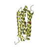
| ||||||||||||||||||||||||||||||||||||||||||||||||||||||||||||||||||||||||||||||||||||||||||||||||||||||||||||||||||||||||||||||||||||||||||||||||||||||
| Unit cell |
| ||||||||||||||||||||||||||||||||||||||||||||||||||||||||||||||||||||||||||||||||||||||||||||||||||||||||||||||||||||||||||||||||||||||||||||||||||||||
| Components on special symmetry positions |
| ||||||||||||||||||||||||||||||||||||||||||||||||||||||||||||||||||||||||||||||||||||||||||||||||||||||||||||||||||||||||||||||||||||||||||||||||||||||
| Noncrystallographic symmetry (NCS) | NCS domain:
NCS domain segments: Component-ID: _ / Beg auth comp-ID: THR / Beg label comp-ID: THR / Refine code: _
NCS ensembles :
|
-Components
| #1: Protein | Mass: 16926.221 Da / Num. of mol.: 4 / Fragment: UNP residues 187-345 Source method: isolated from a genetically manipulated source Source: (gene. exp.) Talaromyces marneffei PM1 (fungus) / Gene: GQ26_0022220 / Plasmid: His-SUMO / Production host:  #2: Chemical | ChemComp-ACD / 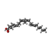  Mass: 304.467 Da / Num. of mol.: 6 / Source method: obtained synthetically / Formula: C20H32O2 Mass: 304.467 Da / Num. of mol.: 6 / Source method: obtained synthetically / Formula: C20H32O2#3: Chemical |   Mass: 92.094 Da / Num. of mol.: 2 / Source method: obtained synthetically / Formula: C3H8O3 Mass: 92.094 Da / Num. of mol.: 2 / Source method: obtained synthetically / Formula: C3H8O3#4: Water | ChemComp-HOH / |  Mass: 18.015 Da / Num. of mol.: 657 / Source method: isolated from a natural source / Formula: H2O Mass: 18.015 Da / Num. of mol.: 657 / Source method: isolated from a natural source / Formula: H2OSequence details | AUTHORS STATE THAT THE GENEBANK ACCESSION NUMBER KFX52825 IS FOR THIS SEQUENCE. | |
|---|
-Experimental details
-Experiment
| Experiment | Method: X-RAY DIFFRACTION / Number of used crystals: 1 |
|---|
- Sample preparation
| Crystal | Density Matthews: 2.14 Å3/Da / Density % sol: 42.54 % |
|---|---|
| Crystal grow | Temperature: 298 K / Method: vapor diffusion, hanging drop / pH: 4.8 / Details: PEG 4000, Sodium Acetate, Ammonium Acetate |
-Data collection
| Diffraction | Mean temperature: 100 K | ||||||||||||||||||||||||||||||||||||||||||||||||||||||||||||||||||
|---|---|---|---|---|---|---|---|---|---|---|---|---|---|---|---|---|---|---|---|---|---|---|---|---|---|---|---|---|---|---|---|---|---|---|---|---|---|---|---|---|---|---|---|---|---|---|---|---|---|---|---|---|---|---|---|---|---|---|---|---|---|---|---|---|---|---|---|
| Diffraction source | Source: SYNCHROTRON / Site: SSRF  / Beamline: BL17U / Wavelength: 0.9793 Å / Beamline: BL17U / Wavelength: 0.9793 Å | ||||||||||||||||||||||||||||||||||||||||||||||||||||||||||||||||||
| Detector | Type: ADSC QUANTUM 315r / Detector: CCD / Date: Mar 26, 2012 | ||||||||||||||||||||||||||||||||||||||||||||||||||||||||||||||||||
| Radiation | Protocol: SINGLE WAVELENGTH / Monochromatic (M) / Laue (L): M / Scattering type: x-ray | ||||||||||||||||||||||||||||||||||||||||||||||||||||||||||||||||||
| Radiation wavelength | Wavelength: 0.9793 Å / Relative weight: 1 | ||||||||||||||||||||||||||||||||||||||||||||||||||||||||||||||||||
| Reflection | Resolution: 1.45→50 Å / Num. obs: 100087 / % possible obs: 99.2 % / Redundancy: 3.9 % / Rmerge(I) obs: 0.087 / Χ2: 1.076 / Net I/av σ(I): 15.889 / Net I/σ(I): 13.8 / Num. measured all: 393148 | ||||||||||||||||||||||||||||||||||||||||||||||||||||||||||||||||||
| Reflection shell | Diffraction-ID: 1 / Rejects: _
|
- Processing
| Software |
| |||||||||||||||||||||||||||||||||||||||||||||||||||||||||||||||||||||||||||||||||||||||||||||||||||||||||||||||||||||||||||||
|---|---|---|---|---|---|---|---|---|---|---|---|---|---|---|---|---|---|---|---|---|---|---|---|---|---|---|---|---|---|---|---|---|---|---|---|---|---|---|---|---|---|---|---|---|---|---|---|---|---|---|---|---|---|---|---|---|---|---|---|---|---|---|---|---|---|---|---|---|---|---|---|---|---|---|---|---|---|---|---|---|---|---|---|---|---|---|---|---|---|---|---|---|---|---|---|---|---|---|---|---|---|---|---|---|---|---|---|---|---|---|---|---|---|---|---|---|---|---|---|---|---|---|---|---|---|---|
| Refinement | Method to determine structure: MOLECULAR REPLACEMENT / Resolution: 1.45→44.99 Å / Cor.coef. Fo:Fc: 0.961 / Cor.coef. Fo:Fc free: 0.951 / WRfactor Rfree: 0.2173 / WRfactor Rwork: 0.1921 / FOM work R set: 0.8634 / SU B: 2.539 / SU ML: 0.049 / SU R Cruickshank DPI: 0.0747 / SU Rfree: 0.0734 / Cross valid method: THROUGHOUT / σ(F): 0 / ESU R: 0.075 / ESU R Free: 0.073 / Stereochemistry target values: MAXIMUM LIKELIHOOD Details: HYDROGENS HAVE BEEN ADDED IN THE RIDING POSITIONS U VALUES : WITH TLS ADDED
| |||||||||||||||||||||||||||||||||||||||||||||||||||||||||||||||||||||||||||||||||||||||||||||||||||||||||||||||||||||||||||||
| Solvent computation | Ion probe radii: 0.8 Å / Shrinkage radii: 0.8 Å / VDW probe radii: 1.2 Å / Solvent model: MASK | |||||||||||||||||||||||||||||||||||||||||||||||||||||||||||||||||||||||||||||||||||||||||||||||||||||||||||||||||||||||||||||
| Displacement parameters | Biso max: 52.51 Å2 / Biso mean: 18.396 Å2 / Biso min: 8.52 Å2
| |||||||||||||||||||||||||||||||||||||||||||||||||||||||||||||||||||||||||||||||||||||||||||||||||||||||||||||||||||||||||||||
| Refinement step | Cycle: final / Resolution: 1.45→44.99 Å
| |||||||||||||||||||||||||||||||||||||||||||||||||||||||||||||||||||||||||||||||||||||||||||||||||||||||||||||||||||||||||||||
| Refine LS restraints |
| |||||||||||||||||||||||||||||||||||||||||||||||||||||||||||||||||||||||||||||||||||||||||||||||||||||||||||||||||||||||||||||
| Refine LS restraints NCS | Refine-ID: X-RAY DIFFRACTION / Type: interatomic distance / Weight position: 0.05
| |||||||||||||||||||||||||||||||||||||||||||||||||||||||||||||||||||||||||||||||||||||||||||||||||||||||||||||||||||||||||||||
| LS refinement shell | Resolution: 1.448→1.486 Å / Total num. of bins used: 20
| |||||||||||||||||||||||||||||||||||||||||||||||||||||||||||||||||||||||||||||||||||||||||||||||||||||||||||||||||||||||||||||
| Refinement TLS params. | Method: refined / Refine-ID: X-RAY DIFFRACTION
| |||||||||||||||||||||||||||||||||||||||||||||||||||||||||||||||||||||||||||||||||||||||||||||||||||||||||||||||||||||||||||||
| Refinement TLS group |
|
