 Movie
Movie Controller

[English] 日本語
 Yorodumi
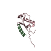
- PDB-6js9: Crystal structure of the N-terminal domain of HtaA from Corynebac... -
+ Open data

- Basic information
| Entry | Database: PDB / ID: 6js9 | ||||||
|---|---|---|---|---|---|---|---|
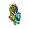
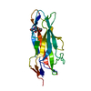
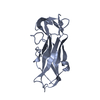
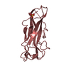
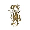
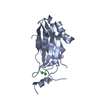
| Title | Crystal structure of the N-terminal domain of HtaA from Corynebacterium glutamicum | ||||||
 Components Components | Hypothetical membrane protein | ||||||
 Keywords Keywords | HEME-BINDING PROTEIN / heme / heme transport | ||||||
| Function / homology | Htaa / Htaa / metal ion binding / PROTOPORPHYRIN IX CONTAINING FE / Hypothetical membrane protein Function and homology information Function and homology information | ||||||
| Biological species |  Corynebacterium glutamicum (bacteria) Corynebacterium glutamicum (bacteria) | ||||||
| Method |  X-RAY DIFFRACTION / SYNCHROTRON / SAD / Resolution: 2 Å X-RAY DIFFRACTION / SYNCHROTRON / SAD / Resolution: 2 Å | ||||||
 Authors Authors | Muraki, N. / Aono, S. | ||||||
| Funding support |  Japan, 1items Japan, 1items
| ||||||
 Citation Citation | Journal: Chem.Commun.(Camb.) / Year: 2019 Title: Structural basis for the heme transfer reaction in heme uptake machinery from Corynebacteria. Authors: Muraki, N. / Kitatsuji, C. / Okamoto, Y. / Uchida, T. / Ishimori, K. / Aono, S. | ||||||
| History |
|
- Structure visualization
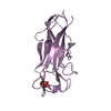
| Structure viewer | Molecule: MolmilJmol/JSmol |
|---|
- Downloads & links
-Download
| PDBx/mmCIF format | 6js9.cif.gz | 150.9 KB | Display | PDBx/mmCIF format |
|---|---|---|---|---|
| PDB format | pdb6js9.ent.gz | 118.9 KB | Display | PDB format |
| PDBx/mmJSON format | 6js9.json.gz | Tree view | PDBx/mmJSON format | |
| Others |  Other downloads Other downloads |
-Validation report
| Arichive directory | https://data.pdbj.org/pub/pdb/validation_reports/js/6js9ftp://data.pdbj.org/pub/pdb/validation_reports/js/6js9 | HTTPS FTP |
|---|
-Related structure data


-Links
 PDBj
PDBj- Assembly
| Deposited unit | 
| ||||||||
|---|---|---|---|---|---|---|---|---|---|
| 1 | 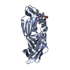
| ||||||||
| 2 | 
| ||||||||
| Unit cell |
|
-Components
| #1: Protein | Mass: 20643.242 Da / Num. of mol.: 2 Source method: isolated from a genetically manipulated source Source: (gene. exp.) Corynebacterium glutamicum (strain ATCC 13032 / DSM 20300 / JCM 1318 / LMG 3730 / NCIMB 10025) (bacteria)Strain: ATCC 13032 / DSM 20300 / JCM 1318 / LMG 3730 / NCIMB 10025 Gene: Cgl0388 / Production host: #2: Chemical |   Mass: 616.487 Da / Num. of mol.: 2 / Source method: obtained synthetically / Formula: C34H32FeN4O4 Mass: 616.487 Da / Num. of mol.: 2 / Source method: obtained synthetically / Formula: C34H32FeN4O4#3: Water | ChemComp-HOH / |  Mass: 18.015 Da / Num. of mol.: 53 / Source method: isolated from a natural source / Formula: H2O Mass: 18.015 Da / Num. of mol.: 53 / Source method: isolated from a natural source / Formula: H2O |
|---|
-Experimental details
-Experiment
| Experiment | Method: X-RAY DIFFRACTION / Number of used crystals: 1 |
|---|
- Sample preparation
| Crystal | Density Matthews: 2.7 Å3/Da / Density % sol: 54.41 % |
|---|---|
| Crystal grow | Temperature: 293 K / Method: vapor diffusion, sitting drop / pH: 8.5 Details: 1.4 M tri-sodium citrate, 0.1 M HEPES NaOH pH7.0, 3% 2-methyl-2,4-pentanediol |
-Data collection
| Diffraction | Mean temperature: 100 K / Serial crystal experiment: N |
|---|---|
| Diffraction source | Source: SYNCHROTRON / Site: SPring-8 / Beamline: BL44XU / Wavelength: 0.9 Å |
| Detector | Type: RAYONIX MX300HE / Detector: CCD / Date: Dec 13, 2014 |
| Radiation | Monochromator: Si (111) double-crystal / Protocol: SINGLE WAVELENGTH / Monochromatic (M) / Laue (L): M / Scattering type: x-ray |
| Radiation wavelength | Wavelength: 0.9 Å / Relative weight: 1 |
| Reflection | Resolution: 2→35.41 Å / Num. obs: 30290 / % possible obs: 99.87 % / Redundancy: 7.1 % / Biso Wilson estimate: 31.65 Å2 / CC1/2: 0.997 / Rmerge(I) obs: 0.0959 / Rpim(I) all: 0.0383 / Rrim(I) all: 0.1035 / Net I/σ(I): 11.01 |
| Reflection shell | Resolution: 2→2.07 Å / Redundancy: 7.1 % / Rmerge(I) obs: 0.6188 / Mean I/σ(I) obs: 3.03 / Num. unique obs: 3011 / CC1/2: 0.792 / Rpim(I) all: 0.2475 / Rrim(I) all: 0.6674 / % possible all: 100 |
- Processing
| Software |
| ||||||||||||||||||||||||||||||||||||||||||||||||||||||||||||||||||||||||||||||||||||
|---|---|---|---|---|---|---|---|---|---|---|---|---|---|---|---|---|---|---|---|---|---|---|---|---|---|---|---|---|---|---|---|---|---|---|---|---|---|---|---|---|---|---|---|---|---|---|---|---|---|---|---|---|---|---|---|---|---|---|---|---|---|---|---|---|---|---|---|---|---|---|---|---|---|---|---|---|---|---|---|---|---|---|---|---|---|
| Refinement | Method to determine structure: SAD / Resolution: 2→35.409 Å / SU ML: 0.25 / Cross valid method: FREE R-VALUE / σ(F): 1.35 / Phase error: 24.55
| ||||||||||||||||||||||||||||||||||||||||||||||||||||||||||||||||||||||||||||||||||||
| Solvent computation | Shrinkage radii: 0.9 Å / VDW probe radii: 1.11 Å | ||||||||||||||||||||||||||||||||||||||||||||||||||||||||||||||||||||||||||||||||||||
| Refinement step | Cycle: LAST / Resolution: 2→35.409 Å
| ||||||||||||||||||||||||||||||||||||||||||||||||||||||||||||||||||||||||||||||||||||
| Refine LS restraints |
| ||||||||||||||||||||||||||||||||||||||||||||||||||||||||||||||||||||||||||||||||||||
| LS refinement shell |
| ||||||||||||||||||||||||||||||||||||||||||||||||||||||||||||||||||||||||||||||||||||
| Refinement TLS params. | Method: refined / Refine-ID: X-RAY DIFFRACTION
| ||||||||||||||||||||||||||||||||||||||||||||||||||||||||||||||||||||||||||||||||||||
| Refinement TLS group |
|
