 Movie
Movie Controller

[English] 日本語
 Yorodumi
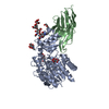
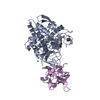
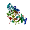
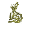
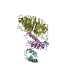
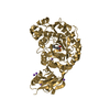
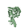
- PDB-5aja: Crystal structure of mandrill SAMHD1 (amino acid residues 1-114) ... -
+ Open data

- Basic information
| Entry | Database: PDB / ID: 5aja | ||||||
|---|---|---|---|---|---|---|---|
| Title | Crystal structure of mandrill SAMHD1 (amino acid residues 1-114) bound to Vpx isolated from mandrill and human DCAF1 (amino acid residues 1058-1396) | ||||||
 Components Components |
| ||||||
 Keywords Keywords | VIRAL PROTEIN / SIV / ACCESSORY PROTEIN / RETROVIRAL RESTRICTION FACTOR / UBIQUITYLATION / PROTEASOMAL DEGRADATION | ||||||
| Function / homology |  Function and homology information Function and homology informationcell competition in a multicellular organism / histone H2AT120 kinase activity / dATP catabolic process / dGTPase activity / dGTP catabolic process / V(D)J recombination / negative regulation of type I interferon-mediated signaling pathway / Cul4-RING E3 ubiquitin ligase complex / ubiquitin-like ligase-substrate adaptor activity / post-translational protein modification ...cell competition in a multicellular organism / histone H2AT120 kinase activity / dATP catabolic process / dGTPase activity / dGTP catabolic process / V(D)J recombination / negative regulation of type I interferon-mediated signaling pathway / Cul4-RING E3 ubiquitin ligase complex / ubiquitin-like ligase-substrate adaptor activity / post-translational protein modification / B cell differentiation / nuclear estrogen receptor binding / virion component / viral penetration into host nucleus / fibrillar center / positive regulation of protein catabolic process / host cell / Antigen processing: Ubiquitination & Proteasome degradation / chromosome / defense response to virus / protein homotetramerization / proteasome-mediated ubiquitin-dependent protein catabolic process / DNA replication / non-specific serine/threonine protein kinase / protein ubiquitination / innate immune response / protein serine kinase activity / DNA repair / symbiont entry into host cell / centrosome / GTP binding / host cell nucleus / negative regulation of transcription by RNA polymerase II / nucleoplasm / ATP binding / metal ion binding / nucleus / cytoplasm Similarity search - Function | ||||||
| Biological species |  HOMO SAPIENS (human) HOMO SAPIENS (human) SIMIAN IMMUNODEFICIENCY VIRUS SIMIAN IMMUNODEFICIENCY VIRUS | ||||||
| Method |  X-RAY DIFFRACTION / SYNCHROTRON / MOLECULAR REPLACEMENT / Resolution: 2.649 Å X-RAY DIFFRACTION / SYNCHROTRON / MOLECULAR REPLACEMENT / Resolution: 2.649 Å | ||||||
 Authors Authors | Schwefel, D. / Boucherit, V.C. / Christodoulou, E. / Walker, P.A. / Stoye, J.P. / Bishop, K.N. / Taylor, I.A. | ||||||
 Citation Citation | Journal: Cell Host Microbe. / Year: 2015 Title: Molecular Determinants for Recognition of Divergent Samhd1 Proteins by the Lentiviral Accessory Protein Vpx. Authors: Schwefel, D. / Boucherit, V.C. / Christodoulou, E. / Walker, P.A. / Stoye, J.P. / Bishop, K.N. / Taylor, I.A. | ||||||
| History |
|
- Structure visualization
| Structure viewer | Molecule: MolmilJmol/JSmol |
|---|
- Downloads & links
-Download
| PDBx/mmCIF format | 5aja.cif.gz | 210.4 KB | Display | PDBx/mmCIF format |
|---|---|---|---|---|
| PDB format | pdb5aja.ent.gz | 167.9 KB | Display | PDB format |
| PDBx/mmJSON format | 5aja.json.gz | Tree view | PDBx/mmJSON format | |
| Others |  Other downloads Other downloads |
-Validation report
| Arichive directory | https://data.pdbj.org/pub/pdb/validation_reports/aj/5ajaftp://data.pdbj.org/pub/pdb/validation_reports/aj/5aja | HTTPS FTP |
|---|
-Related structure data
| Related structure data |  4cc9S S: Starting model for refinement |
|---|---|
| Similar structure data |


-Links
 PDBj
PDBj
- Assembly
| Deposited unit | 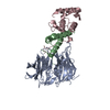
| ||||||||
|---|---|---|---|---|---|---|---|---|---|
| 1 |
| ||||||||
| Unit cell |
|
-Components
| #1: Protein | Mass: 41073.129 Da / Num. of mol.: 1 / Fragment: WD40-REPEAT DOMAIN, RESIDUES 1058-1396 Source method: isolated from a genetically manipulated source Source: (gene. exp.) HOMO SAPIENS (human) / Plasmid: PTRIEX-6 / Cell line (production host): SF9 / Production host:   SPODOPTERA FRUGIPERDA (fall armyworm) SPODOPTERA FRUGIPERDA (fall armyworm)References: UniProt: Q9Y4B6, non-specific serine/threonine protein kinase |
|---|---|
| #2: Protein | Mass: 11940.628 Da / Num. of mol.: 1 Source method: isolated from a genetically manipulated source Source: (gene. exp.) SIMIAN IMMUNODEFICIENCY VIRUS / Strain: MND-2 / Variant: 5440 / Plasmid: PET49B / Production host:  |
| #3: Protein | Mass: 12914.393 Da / Num. of mol.: 1 / Fragment: N-TERMINAL DOMAIN, UNP 1-114 Source method: isolated from a genetically manipulated source Source: (gene. exp.) |
| #4: Chemical | ChemComp-ZN /   Mass: 65.409 Da / Num. of mol.: 1 / Source method: obtained synthetically / Formula: Zn Mass: 65.409 Da / Num. of mol.: 1 / Source method: obtained synthetically / Formula: Zn |
| #5: Water | ChemComp-HOH /  Mass: 18.015 Da / Num. of mol.: 3 / Source method: isolated from a natural source / Formula: H2O Mass: 18.015 Da / Num. of mol.: 3 / Source method: isolated from a natural source / Formula: H2O |
-Experimental details
-Experiment
| Experiment | Method: X-RAY DIFFRACTION / Number of used crystals: 1 |
|---|
- Sample preparation
| Crystal | Density Matthews: 3.04 Å3/Da / Density % sol: 60 % / Description: NONE |
|---|---|
| Crystal grow | Details: 0.16 M TRISODIUM CITRATE-HCL PH 5.2, 4% PEG 6000 |
-Data collection
| Diffraction | Mean temperature: 100 K |
|---|---|
| Diffraction source | Source: SYNCHROTRON / Site: Diamond  / Beamline: I04 / Wavelength: 0.97965 / Beamline: I04 / Wavelength: 0.97965 |
| Detector | Type: DECTRIS PILATUS 6M / Detector: PIXEL / Date: Mar 7, 2014 |
| Radiation | Protocol: SINGLE WAVELENGTH / Monochromatic (M) / Laue (L): M / Scattering type: x-ray |
| Radiation wavelength | Wavelength: 0.97965 Å / Relative weight: 1 |
| Reflection | Resolution: 2.65→30 Å / Num. obs: 24555 / % possible obs: 99.6 % / Observed criterion σ(I): -3 / Redundancy: 9.4 % / Biso Wilson estimate: 76.78 Å2 / Rmerge(I) obs: 0.08 / Net I/σ(I): 20.7 |
| Reflection shell | Resolution: 2.65→2.81 Å / Redundancy: 9.7 % / Rmerge(I) obs: 1.45 / Mean I/σ(I) obs: 1.63 / % possible all: 99.3 |
- Processing
| Software |
| |||||||||||||||||||||||||||||||||||||||||||||||||||||||||||||||||||||||||||||||||||||||||||||||||||||||||||||||||||||||||||||||||||||||||||||||||||||||||||||||||||||||||||||||||||||||||||||||||||||||||||||||||||||||||||||||||||||||||||||||||||||||||||||||||||||||||||||||||||||||||||||||||||||||||||||||||||||||||||||||||||||||||||||||||||||||||||||||||||||||||||||||||||||||
|---|---|---|---|---|---|---|---|---|---|---|---|---|---|---|---|---|---|---|---|---|---|---|---|---|---|---|---|---|---|---|---|---|---|---|---|---|---|---|---|---|---|---|---|---|---|---|---|---|---|---|---|---|---|---|---|---|---|---|---|---|---|---|---|---|---|---|---|---|---|---|---|---|---|---|---|---|---|---|---|---|---|---|---|---|---|---|---|---|---|---|---|---|---|---|---|---|---|---|---|---|---|---|---|---|---|---|---|---|---|---|---|---|---|---|---|---|---|---|---|---|---|---|---|---|---|---|---|---|---|---|---|---|---|---|---|---|---|---|---|---|---|---|---|---|---|---|---|---|---|---|---|---|---|---|---|---|---|---|---|---|---|---|---|---|---|---|---|---|---|---|---|---|---|---|---|---|---|---|---|---|---|---|---|---|---|---|---|---|---|---|---|---|---|---|---|---|---|---|---|---|---|---|---|---|---|---|---|---|---|---|---|---|---|---|---|---|---|---|---|---|---|---|---|---|---|---|---|---|---|---|---|---|---|---|---|---|---|---|---|---|---|---|---|---|---|---|---|---|---|---|---|---|---|---|---|---|---|---|---|---|---|---|---|---|---|---|---|---|---|---|---|---|---|---|---|---|---|---|---|---|---|---|---|---|---|---|---|---|---|---|---|---|---|---|---|---|---|---|---|---|---|---|---|---|---|---|---|---|---|---|---|---|---|---|---|---|---|---|---|---|---|---|---|---|---|---|---|---|---|---|---|---|---|---|---|---|---|---|---|---|---|---|---|---|---|---|---|---|---|---|---|---|---|---|---|---|---|---|---|---|---|---|---|---|---|---|---|---|---|---|---|---|---|---|---|---|
| Refinement | Method to determine structure: MOLECULAR REPLACEMENT Starting model: PDB ENTRY 4CC9 Resolution: 2.649→29.82 Å / SU ML: 0.35 / σ(F): 1.35 / Phase error: 24.27 / Stereochemistry target values: ML
| |||||||||||||||||||||||||||||||||||||||||||||||||||||||||||||||||||||||||||||||||||||||||||||||||||||||||||||||||||||||||||||||||||||||||||||||||||||||||||||||||||||||||||||||||||||||||||||||||||||||||||||||||||||||||||||||||||||||||||||||||||||||||||||||||||||||||||||||||||||||||||||||||||||||||||||||||||||||||||||||||||||||||||||||||||||||||||||||||||||||||||||||||||||||
| Solvent computation | Shrinkage radii: 0.9 Å / VDW probe radii: 1.11 Å / Solvent model: FLAT BULK SOLVENT MODEL | |||||||||||||||||||||||||||||||||||||||||||||||||||||||||||||||||||||||||||||||||||||||||||||||||||||||||||||||||||||||||||||||||||||||||||||||||||||||||||||||||||||||||||||||||||||||||||||||||||||||||||||||||||||||||||||||||||||||||||||||||||||||||||||||||||||||||||||||||||||||||||||||||||||||||||||||||||||||||||||||||||||||||||||||||||||||||||||||||||||||||||||||||||||||
| Displacement parameters | Biso mean: 87.6 Å2 | |||||||||||||||||||||||||||||||||||||||||||||||||||||||||||||||||||||||||||||||||||||||||||||||||||||||||||||||||||||||||||||||||||||||||||||||||||||||||||||||||||||||||||||||||||||||||||||||||||||||||||||||||||||||||||||||||||||||||||||||||||||||||||||||||||||||||||||||||||||||||||||||||||||||||||||||||||||||||||||||||||||||||||||||||||||||||||||||||||||||||||||||||||||||
| Refinement step | Cycle: LAST / Resolution: 2.649→29.82 Å
| |||||||||||||||||||||||||||||||||||||||||||||||||||||||||||||||||||||||||||||||||||||||||||||||||||||||||||||||||||||||||||||||||||||||||||||||||||||||||||||||||||||||||||||||||||||||||||||||||||||||||||||||||||||||||||||||||||||||||||||||||||||||||||||||||||||||||||||||||||||||||||||||||||||||||||||||||||||||||||||||||||||||||||||||||||||||||||||||||||||||||||||||||||||||
| Refine LS restraints |
| |||||||||||||||||||||||||||||||||||||||||||||||||||||||||||||||||||||||||||||||||||||||||||||||||||||||||||||||||||||||||||||||||||||||||||||||||||||||||||||||||||||||||||||||||||||||||||||||||||||||||||||||||||||||||||||||||||||||||||||||||||||||||||||||||||||||||||||||||||||||||||||||||||||||||||||||||||||||||||||||||||||||||||||||||||||||||||||||||||||||||||||||||||||||
| LS refinement shell |
| |||||||||||||||||||||||||||||||||||||||||||||||||||||||||||||||||||||||||||||||||||||||||||||||||||||||||||||||||||||||||||||||||||||||||||||||||||||||||||||||||||||||||||||||||||||||||||||||||||||||||||||||||||||||||||||||||||||||||||||||||||||||||||||||||||||||||||||||||||||||||||||||||||||||||||||||||||||||||||||||||||||||||||||||||||||||||||||||||||||||||||||||||||||||
| Refinement TLS params. | Method: refined / Refine-ID: X-RAY DIFFRACTION
| |||||||||||||||||||||||||||||||||||||||||||||||||||||||||||||||||||||||||||||||||||||||||||||||||||||||||||||||||||||||||||||||||||||||||||||||||||||||||||||||||||||||||||||||||||||||||||||||||||||||||||||||||||||||||||||||||||||||||||||||||||||||||||||||||||||||||||||||||||||||||||||||||||||||||||||||||||||||||||||||||||||||||||||||||||||||||||||||||||||||||||||||||||||||
| Refinement TLS group |
|
