 Movie
Movie Controller

[English] 日本語
 Yorodumi
- PDB-4z8l: Crystal structure of DCAF1/SIV-MND VPX/MND SAMHD1 NTD ternary complex -
+ Open data

- Basic information
| Entry | Database: PDB / ID: 4z8l | ||||||
|---|---|---|---|---|---|---|---|
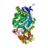
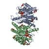
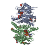
| Title | Crystal structure of DCAF1/SIV-MND VPX/MND SAMHD1 NTD ternary complex | ||||||
 Components Components |
| ||||||
 Keywords Keywords | Viral Protein/VPX-BINDING PROTEIN / HIV / antiviral defense / Viral Protein-VPX-BINDING PROTEIN complex | ||||||
| Function / homology |  Function and homology information Function and homology informationcell competition in a multicellular organism / histone H2AT120 kinase activity / dATP catabolic process / dGTPase activity / dGTP catabolic process / V(D)J recombination / negative regulation of type I interferon-mediated signaling pathway / Cul4-RING E3 ubiquitin ligase complex / ubiquitin-like ligase-substrate adaptor activity / post-translational protein modification ...cell competition in a multicellular organism / histone H2AT120 kinase activity / dATP catabolic process / dGTPase activity / dGTP catabolic process / V(D)J recombination / negative regulation of type I interferon-mediated signaling pathway / Cul4-RING E3 ubiquitin ligase complex / ubiquitin-like ligase-substrate adaptor activity / post-translational protein modification / B cell differentiation / nuclear estrogen receptor binding / virion component / viral penetration into host nucleus / fibrillar center / positive regulation of protein catabolic process / host cell / Antigen processing: Ubiquitination & Proteasome degradation / chromosome / defense response to virus / protein homotetramerization / proteasome-mediated ubiquitin-dependent protein catabolic process / DNA replication / non-specific serine/threonine protein kinase / protein ubiquitination / innate immune response / protein serine kinase activity / DNA repair / symbiont entry into host cell / centrosome / GTP binding / DNA-templated transcription / host cell nucleus / negative regulation of transcription by RNA polymerase II / nucleoplasm / ATP binding / metal ion binding / nucleus / cytoplasm Similarity search - Function | ||||||
| Biological species |  Homo sapiens (human) Homo sapiens (human) Simian immunodeficiency virus Simian immunodeficiency virus | ||||||
| Method |  X-RAY DIFFRACTION / SYNCHROTRON / MOLECULAR REPLACEMENT / Resolution: 2.6 Å X-RAY DIFFRACTION / SYNCHROTRON / MOLECULAR REPLACEMENT / Resolution: 2.6 Å | ||||||
 Authors Authors | Koharudin, L.M. / Wu, Y. / Calero, G. / Ahn, J. / Gronenborn, A.M. | ||||||
| Funding support |  United States, 1items United States, 1items
| ||||||
 Citation Citation | Journal: J.Biol.Chem. / Year: 2015 Title: Structural Basis of Clade-specific Engagement of SAMHD1 (Sterile alpha Motif and Histidine/Aspartate-containing Protein 1) Restriction Factors by Lentiviral Viral Protein X (Vpx) Virulence Factors. Authors: Wu, Y. / Koharudin, L.M. / Mehrens, J. / DeLucia, M. / Byeon, C.H. / Byeon, I.J. / Calero, G. / Ahn, J. / Gronenborn, A.M. | ||||||
| History |
|
- Structure visualization
| Structure viewer | Molecule: MolmilJmol/JSmol |
|---|
- Downloads & links
-Download
| PDBx/mmCIF format | 4z8l.cif.gz | 208.3 KB | Display | PDBx/mmCIF format |
|---|---|---|---|---|
| PDB format | pdb4z8l.ent.gz | 164.6 KB | Display | PDB format |
| PDBx/mmJSON format | 4z8l.json.gz | Tree view | PDBx/mmJSON format | |
| Others |  Other downloads Other downloads |
-Validation report
| Arichive directory | https://data.pdbj.org/pub/pdb/validation_reports/z8/4z8lftp://data.pdbj.org/pub/pdb/validation_reports/z8/4z8l | HTTPS FTP |
|---|
-Related structure data


-Links
 PDBj
PDBj

- Assembly
| Deposited unit | 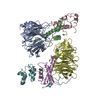
| ||||||||
|---|---|---|---|---|---|---|---|---|---|
| 1 | 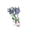
| ||||||||
| 2 | 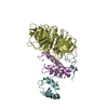
| ||||||||
| Unit cell |
|
-Components
| #1: Protein | Mass: 39777.664 Da / Num. of mol.: 2 / Fragment: UNP residues 1057-1396 Source method: isolated from a genetically manipulated source Source: (gene. exp.) Homo sapiens (human) / Gene: VPRBP, DCAF1, KIAA0800, RIP / Production host: Baculoviridae (virus) / Strain (production host): SF21References: UniProt: Q9Y4B6, non-specific serine/threonine protein kinase #2: Protein | Mass: 12204.781 Da / Num. of mol.: 2 Source method: isolated from a genetically manipulated source Source: (gene. exp.) Simian immunodeficiency virus / Gene: vpx / Plasmid: pET43 / Production host:  #3: Protein | Mass: 13197.734 Da / Num. of mol.: 2 / Fragment: N-terminal Source method: isolated from a genetically manipulated source Source: (gene. exp.) #4: Chemical |   Mass: 65.409 Da / Num. of mol.: 2 / Source method: obtained synthetically / Formula: Zn Mass: 65.409 Da / Num. of mol.: 2 / Source method: obtained synthetically / Formula: Zn#5: Water | ChemComp-HOH / |  Mass: 18.015 Da / Num. of mol.: 114 / Source method: isolated from a natural source / Formula: H2O Mass: 18.015 Da / Num. of mol.: 114 / Source method: isolated from a natural source / Formula: H2O |
|---|
-Experimental details
-Experiment
| Experiment | Method: X-RAY DIFFRACTION |
|---|
- Sample preparation
| Crystal | Density Matthews: 2.2 Å3/Da / Density % sol: 44.07 % |
|---|---|
| Crystal grow | Temperature: 298 K / Method: vapor diffusion, sitting drop / pH: 7.5 Details: 1.6 M NaH2PO4, 0.4 M K2HPO4, 0.1 M Sodium Phosphate/Citrate buffer, pH 4.2 |
-Data collection
| Diffraction | Mean temperature: 100 K |
|---|---|
| Diffraction source | Source: SYNCHROTRON / Site: APS / Beamline: 22-BM / Wavelength: 1 Å |
| Detector | Type: MARMOSAIC 225 mm CCD / Detector: CCD / Date: Jul 25, 2014 |
| Radiation | Monochromator: Rosenbaum-Rock double-crystal Si(220) / Protocol: SINGLE WAVELENGTH / Monochromatic (M) / Laue (L): M / Scattering type: x-ray |
| Radiation wavelength | Wavelength: 1 Å / Relative weight: 1 |
| Reflection | Resolution: 2.6→43.7 Å / Num. obs: 33447 / % possible obs: 98.29 % / Redundancy: 9.1 % / Net I/σ(I): 12.7 |
| Reflection shell | Resolution: 2.6→2.72 Å / % possible obs: 89.3 % / Redundancy: 6.2 % / Rmerge(I) obs: 1.818 / Mean I/σ(I) obs: 1.4 / CC1/2: 0.383 / Rpim(I) all: 0.782 |
- Processing
| Software |
| ||||||||||||||||||||||||||||||||||||||||||||||||||||||||
|---|---|---|---|---|---|---|---|---|---|---|---|---|---|---|---|---|---|---|---|---|---|---|---|---|---|---|---|---|---|---|---|---|---|---|---|---|---|---|---|---|---|---|---|---|---|---|---|---|---|---|---|---|---|---|---|---|---|
| Refinement | Method to determine structure: MOLECULAR REPLACEMENT Starting model: 4CC9, 2E8O Resolution: 2.6→43.695 Å / Cross valid method: FREE R-VALUE / σ(F): 1.92 / Phase error: 25.96 / Stereochemistry target values: TWIN_LSQ_F
| ||||||||||||||||||||||||||||||||||||||||||||||||||||||||
| Solvent computation | Shrinkage radii: 0.9 Å / VDW probe radii: 1.11 Å / Solvent model: FLAT BULK SOLVENT MODEL | ||||||||||||||||||||||||||||||||||||||||||||||||||||||||
| Refinement step | Cycle: LAST / Resolution: 2.6→43.695 Å
| ||||||||||||||||||||||||||||||||||||||||||||||||||||||||
| Refine LS restraints |
| ||||||||||||||||||||||||||||||||||||||||||||||||||||||||
| LS refinement shell |
|
