 Movie
Movie Controller


+ Open data

- Basic information
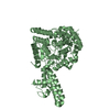
| Entry | Database: PDB / ID: 4xze | ||||||
|---|---|---|---|---|---|---|---|
| Title | The crystal structure of Hazara virus nucleoprotein | ||||||
 Components Components | Nucleoprotein | ||||||
 Keywords Keywords | RNA BINDING PROTEIN / nucleoprotein / Hazara virus | ||||||
| Function / homology |  Function and homology information Function and homology informationhelical viral capsid / viral nucleocapsid / Hydrolases; Acting on ester bonds / ribonucleoprotein complex / hydrolase activity / RNA binding Similarity search - Function | ||||||
| Biological species |  Hazara virus Hazara virus | ||||||
| Method |  X-RAY DIFFRACTION / SYNCHROTRON / MOLECULAR REPLACEMENT / Resolution: 2.9 Å X-RAY DIFFRACTION / SYNCHROTRON / MOLECULAR REPLACEMENT / Resolution: 2.9 Å | ||||||
 Authors Authors | Guo, Y. / Wang, W. / Liu, X. / Wang, X. / Wang, J. / Huo, T. / Liu, B. | ||||||
 Citation Citation | Journal: J.Virol. / Year: 2015 Title: Structural and Functional Diversity of Nairovirus-Encoded Nucleoproteins. Authors: Wang, W. / Liu, X. / Wang, X. / Dong, H. / Ma, C. / Wang, J. / Liu, B. / Mao, Y. / Wang, Y. / Li, T. / Yang, C. / Guo, Y. | ||||||
| History |
|
- Structure visualization
| Structure viewer | Molecule: MolmilJmol/JSmol |
|---|
- Downloads & links
-Download
| PDBx/mmCIF format | 4xze.cif.gz | 369.1 KB | Display | PDBx/mmCIF format |
|---|---|---|---|---|
| PDB format | pdb4xze.ent.gz | 302.9 KB | Display | PDB format |
| PDBx/mmJSON format | 4xze.json.gz | Tree view | PDBx/mmJSON format | |
| Others |  Other downloads Other downloads |
-Validation report
| Arichive directory | https://data.pdbj.org/pub/pdb/validation_reports/xz/4xzeftp://data.pdbj.org/pub/pdb/validation_reports/xz/4xze | HTTPS FTP |
|---|
-Related structure data
| Related structure data |  4xz8C  4xzaC  4xzcC  3u3iS C: citing same article ( S: Starting model for refinement |
|---|---|
| Similar structure data |


-Links
 PDBj
PDBj- Assembly
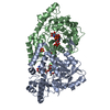
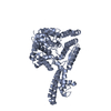
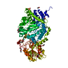
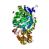
| Deposited unit | 
| ||||||||
|---|---|---|---|---|---|---|---|---|---|
| 1 | 
| ||||||||
| 2 | 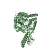
| ||||||||
| 3 | 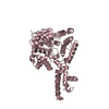
| ||||||||
| 4 | 
| ||||||||
| Unit cell |
|
-Components
| #1: Protein | Mass: 54545.023 Da / Num. of mol.: 4 Source method: isolated from a genetically manipulated source Source: (gene. exp.) Hazara virus / Production host:  #2: Water | ChemComp-HOH / |  Mass: 18.015 Da / Num. of mol.: 104 / Source method: isolated from a natural source / Formula: H2O Mass: 18.015 Da / Num. of mol.: 104 / Source method: isolated from a natural source / Formula: H2O |
|---|
-Experimental details
-Experiment
| Experiment | Method: X-RAY DIFFRACTION |
|---|
- Sample preparation
| Crystal | Density Matthews: 2.53 Å3/Da / Density % sol: 51.35 % |
|---|---|
| Crystal grow | Temperature: 289 K / Method: vapor diffusion, hanging drop / pH: 8 Details: 200mM potassium citrate tribasic monohydrate (PH 8.0), 5mM manganese chloride,14% (wt/vol) PEG 3350 |
-Data collection
| Diffraction | Mean temperature: 100 K |
|---|---|
| Diffraction source | Source: SYNCHROTRON / Site: Photon Factory  / Beamline: BL-17A / Wavelength: 1 Å / Beamline: BL-17A / Wavelength: 1 Å |
| Detector | Type: ADSC QUANTUM 270 / Detector: CCD / Date: May 25, 2014 |
| Radiation | Protocol: SINGLE WAVELENGTH / Monochromatic (M) / Laue (L): M / Scattering type: x-ray |
| Radiation wavelength | Wavelength: 1 Å / Relative weight: 1 |
| Reflection | Resolution: 2.9→50 Å / Num. obs: 42788 / % possible obs: 84.7 % / Redundancy: 6 % / Net I/σ(I): 11.008 |
| Reflection shell | Resolution: 2.9→2.95 Å / Redundancy: 7.1 % / Rmerge(I) obs: 0.352 / Mean I/σ(I) obs: 0.327 / % possible all: 100 |
- Processing
| Software |
| ||||||||||||||||||||||||||||||||||||||||||||||||||||||||||||||||||||||||||||||||||||||||||||||||||||||||||||||||
|---|---|---|---|---|---|---|---|---|---|---|---|---|---|---|---|---|---|---|---|---|---|---|---|---|---|---|---|---|---|---|---|---|---|---|---|---|---|---|---|---|---|---|---|---|---|---|---|---|---|---|---|---|---|---|---|---|---|---|---|---|---|---|---|---|---|---|---|---|---|---|---|---|---|---|---|---|---|---|---|---|---|---|---|---|---|---|---|---|---|---|---|---|---|---|---|---|---|---|---|---|---|---|---|---|---|---|---|---|---|---|---|---|---|
| Refinement | Method to determine structure: MOLECULAR REPLACEMENT Starting model: 3U3I Resolution: 2.9→48.22 Å / SU ML: 0.39 / Cross valid method: FREE R-VALUE / σ(F): 1.35 / Phase error: 26.91 / Stereochemistry target values: ML
| ||||||||||||||||||||||||||||||||||||||||||||||||||||||||||||||||||||||||||||||||||||||||||||||||||||||||||||||||
| Solvent computation | Shrinkage radii: 1.06 Å / VDW probe radii: 1.3 Å / Solvent model: FLAT BULK SOLVENT MODEL / Bsol: 45.18 Å2 / ksol: 0.35 e/Å3 | ||||||||||||||||||||||||||||||||||||||||||||||||||||||||||||||||||||||||||||||||||||||||||||||||||||||||||||||||
| Displacement parameters |
| ||||||||||||||||||||||||||||||||||||||||||||||||||||||||||||||||||||||||||||||||||||||||||||||||||||||||||||||||
| Refinement step | Cycle: LAST / Resolution: 2.9→48.22 Å
| ||||||||||||||||||||||||||||||||||||||||||||||||||||||||||||||||||||||||||||||||||||||||||||||||||||||||||||||||
| Refine LS restraints |
| ||||||||||||||||||||||||||||||||||||||||||||||||||||||||||||||||||||||||||||||||||||||||||||||||||||||||||||||||
| LS refinement shell |
|
