PAS domain / PAS-associated, C-terminal / PAC domain profile. / PAC motif / Motif C-terminal to PAS motifs (likely to contribute to PAS structural domain) / PAS domain / Beta-Lactamase / PAS repeat profile. / PAS domain / PAS domain superfamily ...PAS domain / PAS-associated, C-terminal / PAC domain profile. / PAC motif / Motif C-terminal to PAS motifs (likely to contribute to PAS structural domain) / PAS domain / Beta-Lactamase / PAS repeat profile. / PAS domain / PAS domain superfamily / 2-Layer Sandwich / Alpha Beta Similarity search - Domain/homology
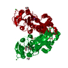
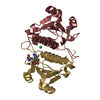
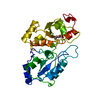
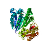
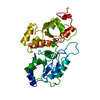
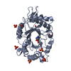
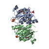
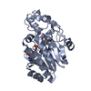
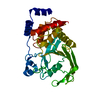
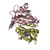
Journal: Structure / Year: 2016 Title: Structure of a Native-like Aureochrome 1a LOV Domain Dimer from Phaeodactylum tricornutum. Authors: Ankan Banerjee / Elena Herman / Tilman Kottke / Lars-Oliver Essen / Abstract: Light-oxygen-voltage (LOV) domains absorb blue light for mediating various biological responses in all three domains of life. Aureochromes from stramenopile algae represent a subfamily of ...Light-oxygen-voltage (LOV) domains absorb blue light for mediating various biological responses in all three domains of life. Aureochromes from stramenopile algae represent a subfamily of photoreceptors that differs by its inversed topology with a C-terminal LOV sensor and an N-terminal effector (basic region leucine zipper, bZIP) domain. We crystallized the LOV domain including its flanking helices, A'α and Jα, of aureochrome 1a from Phaeodactylum tricornutum in the dark state and solved the structure at 2.8 Å resolution. Both flanking helices contribute to the interface of the native-like dimer. Small-angle X-ray scattering shows light-induced conformational changes limited to the dimeric envelope as well as increased flexibility in the lit state for the flanking helices. These rearrangements are considered to be crucial for the formation of the light-activated dimer. Finally, the LOV domain of the class 2 aureochrome PtAUREO2 was shown to lack a chromophore because of steric hindrance caused by M301.
In the structure databanks used in Yorodumi, some data are registered as the other names, "COVID-19 virus" and "2019-nCoV". Here are the details of the virus and the list of structure data.
Jan 31, 2019. EMDB accession codes are about to change! (news from PDBe EMDB page)
EMDB accession codes are about to change! (news from PDBe EMDB page)
The allocation of 4 digits for EMDB accession codes will soon come to an end. Whilst these codes will remain in use, new EMDB accession codes will include an additional digit and will expand incrementally as the available range of codes is exhausted. The current 4-digit format prefixed with “EMD-” (i.e. EMD-XXXX) will advance to a 5-digit format (i.e. EMD-XXXXX), and so on. It is currently estimated that the 4-digit codes will be depleted around Spring 2019, at which point the 5-digit format will come into force.
The EM Navigator/Yorodumi systems omit the EMD- prefix.
Related info.:Q: What is EMD? / ID/Accession-code notation in Yorodumi/EM Navigator
Yorodumi is a browser for structure data from EMDB, PDB, SASBDB, etc.
This page is also the successor to EM Navigator detail page, and also detail information page/front-end page for Omokage search.
The word "yorodu" (or yorozu) is an old Japanese word meaning "ten thousand". "mi" (miru) is to see.
Related info.:EMDB / PDB / SASBDB / Comparison of 3 databanks / Yorodumi Search / Aug 31, 2016. New EM Navigator & Yorodumi / Yorodumi Papers / Jmol/JSmol / Function and homology information / Changes in new EM Navigator and Yorodumi
 Movie
Movie Controller

 Yorodumi
 Open data
 Basic information
 Components
 Keywords
 Function and homology information
 X-RAY DIFFRACTION /
  Authors
 Citation
 Structure visualization
 Downloads & links
 Other downloads
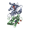


 PDBj
PDBj


 Assembly
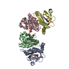


 Mass: 456.344 Da / Num. of mol.: 4 / Source method: obtained synthetically / Formula: C17H21N4O9P
 Mass: 35.453 Da / Num. of mol.: 2 / Source method: obtained synthetically / Formula: Cl
 Mass: 92.094 Da / Num. of mol.: 1 / Source method: obtained synthetically / Formula: C3H8O3
 Mass: 18.015 Da / Num. of mol.: 32 / Source method: isolated from a natural source / Formula: H2O
 Sample preparation
 Processing