 Movie
Movie Controller

[English] 日本語
 Yorodumi
- PDB-5a8j: Crystal structure of the ArnB paralog VWA2 from Sulfolobus acidoc... -
+ Open data

- Basic information
| Entry | Database: PDB / ID: 5a8j | ||||||
|---|---|---|---|---|---|---|---|
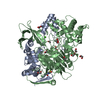
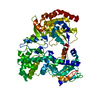
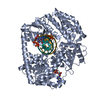
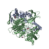
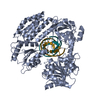
| Title | Crystal structure of the ArnB paralog VWA2 from Sulfolobus acidocaldarius | ||||||
 Components Components | VWA2 | ||||||
 Keywords Keywords | TRANSCRIPTION / VON WILLEBRAND / PHOSPHORYLATION | ||||||
| Function / homology | Archaellum regulatory network B, C-terminal / Archaellum regulatory network B, C-terminal domain / von Willebrand factor type A domain / VWFA domain profile. / von Willebrand factor (vWF) type A domain / von Willebrand factor, type A / von Willebrand factor A-like domain superfamily / CITRIC ACID / Conserved protein Function and homology information Function and homology information | ||||||
| Biological species |   SULFOLOBUS ACIDOCALDARIUS (acidophilic) SULFOLOBUS ACIDOCALDARIUS (acidophilic) | ||||||
| Method |  X-RAY DIFFRACTION / SYNCHROTRON / SAD / Resolution: 1.46 Å X-RAY DIFFRACTION / SYNCHROTRON / SAD / Resolution: 1.46 Å | ||||||
 Authors Authors | Hoffmann, L. / Anders, K. / Reimann, J. / Linne, U. / Essen, L.-O. / Albers, S.-V. | ||||||
 Citation Citation | Journal: To be Published Title: Phosphorylation-Dependent Interaction between an Archaeal Von Willebrand and Fha Domain Recruits Arna-Arnb Complex to DNA for Repression of Motility Authors: Hoffmann, L. / Anders, K. / Reimann, J. / Linne, U. / Essen, L.-O. / Albers, S.-V. | ||||||
| History |
|
- Structure visualization
| Structure viewer | Molecule: MolmilJmol/JSmol |
|---|
- Downloads & links
-Download
| PDBx/mmCIF format | 5a8j.cif.gz | 233.2 KB | Display | PDBx/mmCIF format |
|---|---|---|---|---|
| PDB format | pdb5a8j.ent.gz | 192.9 KB | Display | PDB format |
| PDBx/mmJSON format | 5a8j.json.gz | Tree view | PDBx/mmJSON format | |
| Others |  Other downloads Other downloads |
-Validation report
| Arichive directory | https://data.pdbj.org/pub/pdb/validation_reports/a8/5a8jftp://data.pdbj.org/pub/pdb/validation_reports/a8/5a8j | HTTPS FTP |
|---|
-Related structure data


-Links
 PDBj
PDBj


- Assembly
| Deposited unit | 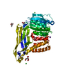
| ||||||||
|---|---|---|---|---|---|---|---|---|---|
| 1 | 
| ||||||||
| Unit cell |
| ||||||||
| Components on special symmetry positions |
|
-Components
-Protein , 1 types, 1 molecules A
| #1: Protein | Mass: 41772.461 Da / Num. of mol.: 1 / Fragment: VON WILLEBRAND DOMAIN, RESIDUES 2-360 Source method: isolated from a genetically manipulated source Source: (gene. exp.) SULFOLOBUS ACIDOCALDARIUS (acidophilic)Strain: DSM639 / Production host:  |
|---|
-Non-polymers , 6 types, 332 molecules 


| #2: Chemical | ChemComp-SO4 /  Mass: 96.063 Da / Num. of mol.: 1 / Source method: obtained synthetically / Formula: SO4 Mass: 96.063 Da / Num. of mol.: 1 / Source method: obtained synthetically / Formula: SO4 | ||||||||
|---|---|---|---|---|---|---|---|---|---|
| #3: Chemical | ChemComp-GOL /  Mass: 92.094 Da / Num. of mol.: 4 / Source method: obtained synthetically / Formula: C3H8O3 Mass: 92.094 Da / Num. of mol.: 4 / Source method: obtained synthetically / Formula: C3H8O3#4: Chemical |  Mass: 35.453 Da / Num. of mol.: 2 / Source method: obtained synthetically / Formula: Cl Mass: 35.453 Da / Num. of mol.: 2 / Source method: obtained synthetically / Formula: Cl#5: Chemical | ChemComp-CIT / |  Mass: 192.124 Da / Num. of mol.: 1 / Source method: obtained synthetically / Formula: C6H8O7 Mass: 192.124 Da / Num. of mol.: 1 / Source method: obtained synthetically / Formula: C6H8O7#6: Chemical | ChemComp-NA / |  Mass: 22.990 Da / Num. of mol.: 1 / Source method: obtained synthetically / Formula: Na Mass: 22.990 Da / Num. of mol.: 1 / Source method: obtained synthetically / Formula: Na#7: Water | ChemComp-HOH / | Mass: 18.015 Da / Num. of mol.: 323 / Source method: isolated from a natural source / Formula: H2O |
-Experimental details
-Experiment
| Experiment | Method: X-RAY DIFFRACTION |
|---|
- Sample preparation
| Crystal | Density Matthews: 2.26 Å3/Da / Density % sol: 45.46 % / Description: NONE |
|---|---|
| Crystal grow | Details: RESERVOIR BUFFER: 0.1 M SODIUMCITRATE PH 5.5; 2.5 M AMMONIUMSULFATE; PROTEIN BUFFER: 20 MM HEPES PH 7.0; 100 MM KCL, 2.7 MG/ML PROTEIN IN PROTEIN BUFFER; MIX RESERVOIR AND PROTEIN 1:1 |
-Data collection
| Diffraction | Mean temperature: 100 K |
|---|---|
| Diffraction source | Source: SYNCHROTRON / Site: ESRF  / Beamline: ID29 / Wavelength: 0.70847 / Beamline: ID29 / Wavelength: 0.70847 |
| Detector | Type: DECTRIS PILATUS 6M / Detector: PIXEL / Date: Jun 10, 2011 |
| Radiation | Monochromator: SI(311) / Protocol: SINGLE WAVELENGTH / Monochromatic (M) / Laue (L): M / Scattering type: x-ray |
| Radiation wavelength | Wavelength: 0.70847 Å / Relative weight: 1 |
| Reflection | Resolution: 1.46→41.66 Å / Num. obs: 65094 / % possible obs: 99.2 % / Observed criterion σ(I): -3 / Redundancy: 5.4 % / Biso Wilson estimate: 16.79 Å2 / Rmerge(I) obs: 0.05 / Net I/σ(I): 17.5 |
| Reflection shell | Resolution: 1.46→1.54 Å / Redundancy: 5.6 % / Rmerge(I) obs: 0.49 / Mean I/σ(I) obs: 3.5 / % possible all: 97.7 |
- Processing
| Software |
| |||||||||||||||||||||||||||||||||||||||||||||||||||||||||||||||||||||||||||||
|---|---|---|---|---|---|---|---|---|---|---|---|---|---|---|---|---|---|---|---|---|---|---|---|---|---|---|---|---|---|---|---|---|---|---|---|---|---|---|---|---|---|---|---|---|---|---|---|---|---|---|---|---|---|---|---|---|---|---|---|---|---|---|---|---|---|---|---|---|---|---|---|---|---|---|---|---|---|---|
| Refinement | Method to determine structure: SAD Starting model: NONE Resolution: 1.46→41.658 Å / SU ML: 0.18 / σ(F): 1.37 / Phase error: 21.96 / Stereochemistry target values: ML Details: RESIDUES A2 - G366 OF VWA2 CAN BE SEEN IN THE STRUCTURE
| |||||||||||||||||||||||||||||||||||||||||||||||||||||||||||||||||||||||||||||
| Solvent computation | Shrinkage radii: 1.1 Å / VDW probe radii: 1.3 Å / Solvent model: FLAT BULK SOLVENT MODEL | |||||||||||||||||||||||||||||||||||||||||||||||||||||||||||||||||||||||||||||
| Displacement parameters | Biso mean: 19 Å2 | |||||||||||||||||||||||||||||||||||||||||||||||||||||||||||||||||||||||||||||
| Refinement step | Cycle: LAST / Resolution: 1.46→41.658 Å
| |||||||||||||||||||||||||||||||||||||||||||||||||||||||||||||||||||||||||||||
| Refine LS restraints |
| |||||||||||||||||||||||||||||||||||||||||||||||||||||||||||||||||||||||||||||
| LS refinement shell |
|
