 Movie
Movie Controller

[English] 日本語
 Yorodumi
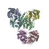
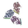
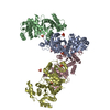
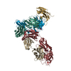
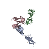
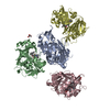
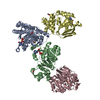
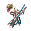
- PDB-5a5u: Structure of mammalian eIF3 in the context of the 43S preinitiati... -
+ Open data

- Basic information
| Entry | Database: PDB / ID: 5a5u | ||||||
|---|---|---|---|---|---|---|---|
| Title | Structure of mammalian eIF3 in the context of the 43S preinitiation complex | ||||||
 Components Components |
| ||||||
 Keywords Keywords | TRANSLATION / EIF3 / EIF3 OCTAMER CORE / MAMMALIAN PREINITIATION 34S COMPLEX / EIF3G/I/B / EIF3D | ||||||
| Function / homology |  Function and homology information Function and homology informationviral translational termination-reinitiation / eukaryotic translation initiation factor 3 complex, eIF3m / IRES-dependent viral translational initiation / translation reinitiation / formation of cytoplasmic translation initiation complex / multi-eIF complex / eukaryotic translation initiation factor 3 complex / eukaryotic 43S preinitiation complex / eukaryotic 48S preinitiation complex / regulation of translational initiation ...viral translational termination-reinitiation / eukaryotic translation initiation factor 3 complex, eIF3m / IRES-dependent viral translational initiation / translation reinitiation / formation of cytoplasmic translation initiation complex / multi-eIF complex / eukaryotic translation initiation factor 3 complex / eukaryotic 43S preinitiation complex / eukaryotic 48S preinitiation complex / regulation of translational initiation / Formation of the ternary complex, and subsequently, the 43S complex / Translation initiation complex formation / Ribosomal scanning and start codon recognition / cytoplasmic translational initiation / L13a-mediated translational silencing of Ceruloplasmin expression / translation initiation factor binding / translation initiation factor activity / translational initiation / cytoplasmic stress granule / RNA binding Similarity search - Function | ||||||
| Biological species |  | ||||||
| Method | ELECTRON MICROSCOPY / single particle reconstruction / cryo EM / Resolution: 9 Å | ||||||
 Authors Authors | des-Georges, A. / Dhote, V. / Kuhn, L. / Hellen, C.U.T. / Pestova, T.V. / Frank, J. / Hashem, Y. | ||||||
 Citation Citation | Journal: Nature / Year: 2015 Title: Structure of mammalian eIF3 in the context of the 43S preinitiation complex. Authors: Amedee des Georges / Vidya Dhote / Lauriane Kuhn / Christopher U T Hellen / Tatyana V Pestova / Joachim Frank / Yaser Hashem /   Abstract: During eukaryotic translation initiation, 43S complexes, comprising a 40S ribosomal subunit, initiator transfer RNA and initiation factors (eIF) 2, 3, 1 and 1A, attach to the 5'-terminal region of ...During eukaryotic translation initiation, 43S complexes, comprising a 40S ribosomal subunit, initiator transfer RNA and initiation factors (eIF) 2, 3, 1 and 1A, attach to the 5'-terminal region of messenger RNA and scan along it to the initiation codon. Scanning on structured mRNAs also requires the DExH-box protein DHX29. Mammalian eIF3 contains 13 subunits and participates in nearly all steps of translation initiation. Eight subunits having PCI (proteasome, COP9 signalosome, eIF3) or MPN (Mpr1, Pad1, amino-terminal) domains constitute the structural core of eIF3, to which five peripheral subunits are flexibly linked. Here we present a cryo-electron microscopy structure of eIF3 in the context of the DHX29-bound 43S complex, showing the PCI/MPN core at ∼6 Å resolution. It reveals the organization of the individual subunits and their interactions with components of the 43S complex. We were able to build near-complete polyalanine-level models of the eIF3 PCI/MPN core and of two peripheral subunits. The implications for understanding mRNA ribosomal attachment and scanning are discussed. | ||||||
| History |
|
- Structure visualization
| Movie |
Movie viewer |
|---|---|
| Structure viewer | Molecule: MolmilJmol/JSmol |
- Downloads & links
-Download
| PDBx/mmCIF format | 5a5u.cif.gz | 208.2 KB | Display | PDBx/mmCIF format |
|---|---|---|---|---|
| PDB format | pdb5a5u.ent.gz | 154 KB | Display | PDB format |
| PDBx/mmJSON format | 5a5u.json.gz | Tree view | PDBx/mmJSON format | |
| Others |  Other downloads Other downloads |
-Validation report
| Arichive directory | https://data.pdbj.org/pub/pdb/validation_reports/a5/5a5uftp://data.pdbj.org/pub/pdb/validation_reports/a5/5a5u | HTTPS FTP |
|---|
-Related structure data
| Related structure data | 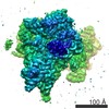 3057MC 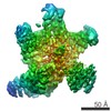 3056C 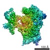 3058C 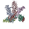 5a5tC C: citing same article ( M: map data used to model this data |
|---|---|
| Similar structure data |
-Links
 PDBj
PDBj


- Assembly
| Deposited unit | 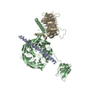
|
|---|---|
| 1 |
|
-Components
| #1: Protein | Mass: 4613.678 Da / Num. of mol.: 1 / Source method: isolated from a natural source / Source: (natural) |
|---|---|
| #2: Protein | Mass: 124402.336 Da / Num. of mol.: 1 / Source method: isolated from a natural source / Source: (natural) |
| #3: Protein | Mass: 38803.375 Da / Num. of mol.: 1 / Source method: isolated from a natural source / Source: (natural) |
| Sequence details | THE SAMPLE OF CHAIN B CONTAINS FULL LENGTH EIF3B. |
-Experimental details
-Experiment
| Experiment | Method: ELECTRON MICROSCOPY |
|---|---|
| EM experiment | Aggregation state: PARTICLE / 3D reconstruction method: single particle reconstruction |
- Sample preparation
| Component | Name: MAMMALIAN 43S PREINITIATION COMPLEX BOUND TO DHX29 / Type: RIBOSOME |
|---|---|
| Specimen | Embedding applied: NO / Shadowing applied: NO / Staining applied: NO / Vitrification applied: YES |
| Specimen support | Details: CARBON |
| Vitrification | Instrument: FEI VITROBOT MARK IV / Cryogen name: ETHANE Details: VITRIFICATION 1 -- CRYOGEN- ETHANE, HUMIDITY- 100, TEMPERATURE- 120, INSTRUMENT- FEI VITROBOT MARK IV, |
- Electron microscopy imaging
| Microscopy | Model: FEI/PHILIPS CM300FEG/HE / Date: Nov 1, 2013 |
|---|---|
| Electron gun | Electron source:  FIELD EMISSION GUN / Accelerating voltage: 300 kV / Illumination mode: FLOOD BEAM FIELD EMISSION GUN / Accelerating voltage: 300 kV / Illumination mode: FLOOD BEAM |
| Electron lens | Mode: BRIGHT FIELD / Calibrated magnification: 30120 X / Nominal defocus max: 4500 nm / Nominal defocus min: 500 nm / Cs: 2 mm |
| Specimen holder | Temperature: 100 K |
| Image recording | Electron dose: 25 e/Å2 / Film or detector model: GATAN K2 (4k x 4k) |
- Processing
| EM software | Name: RELION / Category: 3D reconstruction | ||||||||||||
|---|---|---|---|---|---|---|---|---|---|---|---|---|---|
| CTF correction | Details: EACH PARTICLE | ||||||||||||
| Symmetry | Point symmetry: C1 (asymmetric) | ||||||||||||
| 3D reconstruction | Method: TEMPLATE MATCHING / Resolution: 9 Å / Num. of particles: 54410 Details: SUBMISSION BASED ON EXPERIMENTAL DATA FROM EMDB EMD-3057. Symmetry type: POINT | ||||||||||||
| Refinement | Highest resolution: 9 Å | ||||||||||||
| Refinement step | Cycle: LAST / Highest resolution: 9 Å
|
