 ムービー
ムービー コントローラー


+ データを開く

- 基本情報
基本情報
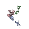
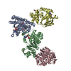
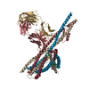
| 登録情報 | データベース: PDB / ID: 5a5u | ||||||
|---|---|---|---|---|---|---|---|
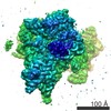
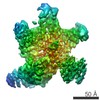
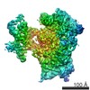
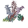
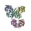
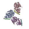
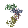
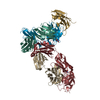
| タイトル | Structure of mammalian eIF3 in the context of the 43S preinitiation complex | ||||||
 要素 要素 |
| ||||||
 キーワード キーワード |  TRANSLATION (翻訳 (生物学)) / EIF3 (EIF3) / EIF3 OCTAMER CORE / MAMMALIAN PREINITIATION 34S COMPLEX / EIF3G/I/B / EIF3D TRANSLATION (翻訳 (生物学)) / EIF3 (EIF3) / EIF3 OCTAMER CORE / MAMMALIAN PREINITIATION 34S COMPLEX / EIF3G/I/B / EIF3D | ||||||
| 機能・相同性 |  機能・相同性情報 機能・相同性情報viral translational termination-reinitiation / eukaryotic translation initiation factor 3 complex, eIF3m / translation reinitiation / IRES-dependent viral translational initiation / multi-eIF complex / eukaryotic translation initiation factor 3 complex / eukaryotic 43S preinitiation complex / cytoplasmic translational initiation / formation of cytoplasmic translation initiation complex / eukaryotic 48S preinitiation complex ...viral translational termination-reinitiation / eukaryotic translation initiation factor 3 complex, eIF3m / translation reinitiation / IRES-dependent viral translational initiation / multi-eIF complex / eukaryotic translation initiation factor 3 complex / eukaryotic 43S preinitiation complex / cytoplasmic translational initiation / formation of cytoplasmic translation initiation complex / eukaryotic 48S preinitiation complex / regulation of translational initiation / Formation of the ternary complex, and subsequently, the 43S complex / Translation initiation complex formation / Ribosomal scanning and start codon recognition / L13a-mediated translational silencing of Ceruloplasmin expression / translation initiation factor binding / translational initiation / translation initiation factor activity / cytoplasmic stress granule / RNA binding類似検索 - 分子機能 | ||||||
| 生物種 |  ORYCTOLAGUS CUNICULUS (ウサギ) ORYCTOLAGUS CUNICULUS (ウサギ) | ||||||
| 手法 | 電子顕微鏡法 / 単粒子再構成法 / クライオ電子顕微鏡法 / 解像度: 9 Å | ||||||
 データ登録者 データ登録者 | des-Georges, A. / Dhote, V. / Kuhn, L. / Hellen, C.U.T. / Pestova, T.V. / Frank, J. / Hashem, Y. | ||||||
 引用 引用 | ジャーナル: Nature / 年: 2015 タイトル: Structure of mammalian eIF3 in the context of the 43S preinitiation complex. 著者: Amedee des Georges / Vidya Dhote / Lauriane Kuhn / Christopher U T Hellen / Tatyana V Pestova / Joachim Frank / Yaser Hashem /   要旨: During eukaryotic translation initiation, 43S complexes, comprising a 40S ribosomal subunit, initiator transfer RNA and initiation factors (eIF) 2, 3, 1 and 1A, attach to the 5'-terminal region of ...During eukaryotic translation initiation, 43S complexes, comprising a 40S ribosomal subunit, initiator transfer RNA and initiation factors (eIF) 2, 3, 1 and 1A, attach to the 5'-terminal region of messenger RNA and scan along it to the initiation codon. Scanning on structured mRNAs also requires the DExH-box protein DHX29. Mammalian eIF3 contains 13 subunits and participates in nearly all steps of translation initiation. Eight subunits having PCI (proteasome, COP9 signalosome, eIF3) or MPN (Mpr1, Pad1, amino-terminal) domains constitute the structural core of eIF3, to which five peripheral subunits are flexibly linked. Here we present a cryo-electron microscopy structure of eIF3 in the context of the DHX29-bound 43S complex, showing the PCI/MPN core at ∼6 Å resolution. It reveals the organization of the individual subunits and their interactions with components of the 43S complex. We were able to build near-complete polyalanine-level models of the eIF3 PCI/MPN core and of two peripheral subunits. The implications for understanding mRNA ribosomal attachment and scanning are discussed. | ||||||
| 履歴 |
|
- 構造の表示
構造の表示
| ムービー |
ムービービューア |
|---|---|
| 構造ビューア | 分子: MolmilJmol/JSmol |
- ダウンロードとリンク
-ダウンロード
| PDBx/mmCIF形式 | 5a5u.cif.gz | 204.5 KB | 表示 | PDBx/mmCIF形式 |
|---|---|---|---|---|
| PDB形式 | pdb5a5u.ent.gz | 157.9 KB | 表示 | PDB形式 |
| PDBx/mmJSON形式 | 5a5u.json.gz | ツリー表示 | PDBx/mmJSON形式 | |
| その他 |  その他のダウンロード その他のダウンロード |
-検証レポート
| アーカイブディレクトリ | https://data.pdbj.org/pub/pdb/validation_reports/a5/5a5uftp://data.pdbj.org/pub/pdb/validation_reports/a5/5a5u | HTTPS FTP |
|---|
-関連構造データ
-リンク
 PDBj
PDBj


- 集合体
集合体
| 登録構造単位 | 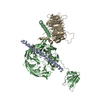
|
|---|---|
| 1 |
|
-要素
| #1: タンパク質 | EIF3 分子量: 4613.678 Da / 分子数: 1 / 由来タイプ: 天然 / 由来: (天然) ORYCTOLAGUS CUNICULUS (ウサギ) / Cell: RETICULCYTES / 組織: BLOOD血液 |
|---|---|
| #2: タンパク質 | EIF3 / EUKARYOTIC INITIATION FACTOR 3 分子量: 124402.336 Da / 分子数: 1 / 由来タイプ: 天然 / 由来: (天然) ORYCTOLAGUS CUNICULUS (ウサギ) / Cell: RETICULCYTES / 組織: BLOOD血液 / 参照: UniProt: G1SZ03 |
| #3: タンパク質 | EIF3 / EIF3I / EUKARYOTIC TRANSLATION INITIATION FACTOR 3 39 KDA SUBUNIT / EIF-3 39 KDA SUBUNIT / EIF3 P39 ...EIF3I / EUKARYOTIC TRANSLATION INITIATION FACTOR 3 39 KDA SUBUNIT / EIF-3 39 KDA SUBUNIT / EIF3 P39 / EUKARYOTIC INITIATION FACTOR 3 分子量: 38803.375 Da / 分子数: 1 / 由来タイプ: 天然 / 由来: (天然) ORYCTOLAGUS CUNICULUS (ウサギ) / Cell: RETICULCYTES / 組織: BLOOD血液 / 参照: UniProt: P40217 |
| 配列の詳細 | THE SAMPLE OF CHAIN B CONTAINS FULL LENGTH EIF3B. |
-実験情報
-実験
| 実験 | 手法: 電子顕微鏡法 |
|---|---|
| EM実験 | 試料の集合状態: PARTICLE / 3次元再構成法: 単粒子再構成法 |
- 試料調製
試料調製
| 構成要素 | 名称: MAMMALIAN 43S PREINITIATION COMPLEX BOUND TO DHX29 / タイプ: RIBOSOME |
|---|---|
| 試料 | 包埋: NO / シャドウイング: NO / 染色: NO / 凍結: YES |
| 試料支持 | 詳細: CARBON |
| 急速凍結 | 装置: FEI VITROBOT MARK IV / 凍結剤: ETHANE 詳細: VITRIFICATION 1 -- CRYOGEN- ETHANE, HUMIDITY- 100, TEMPERATURE- 120, INSTRUMENT- FEI VITROBOT MARK IV, |
- 電子顕微鏡撮影
| 顕微鏡 | モデル: FEI/PHILIPS CM300FEG/HE / 日付: 2013年11月1日 |
|---|---|
| 電子銃 | 電子線源: FIELD EMISSION GUN / 加速電圧: 300 kV / 照射モード: FLOOD BEAM |
| 電子レンズ | モード: BRIGHT FIELDBright-field microscopy / 倍率(補正後): 30120 X / 最大 デフォーカス(公称値): 4500 nm / 最小 デフォーカス(公称値): 500 nm / Cs: 2 mm |
| 試料ホルダ | 温度: 100 K |
| 撮影 | 電子線照射量: 25 e/Å2 / フィルム・検出器のモデル: GATAN K2 (4k x 4k) |
- 解析
解析
| EMソフトウェア | 名称: RELION / カテゴリ: 3次元再構成 | ||||||||||||
|---|---|---|---|---|---|---|---|---|---|---|---|---|---|
| CTF補正 | 詳細: EACH PARTICLE | ||||||||||||
| 対称性 | 点対称性: C1 (非対称) | ||||||||||||
| 3次元再構成 | 手法: TEMPLATE MATCHING / 解像度: 9 Å / 粒子像の数: 54410 詳細: SUBMISSION BASED ON EXPERIMENTAL DATA FROM EMDB EMD-3057. 対称性のタイプ: POINT | ||||||||||||
| 精密化 | 最高解像度: 9 Å | ||||||||||||
| 精密化ステップ | サイクル: LAST / 最高解像度: 9 Å
|
