 Movie
Movie Controller

[English] 日本語
 Yorodumi
- EMDB-3058: Structure of mammalian eIF3 in the context of the 43S preinitiati... -
+ Open data

- Basic information
| Entry | Database: EMDB / ID: EMD-3058 | |||||||||
|---|---|---|---|---|---|---|---|---|---|---|
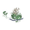
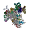
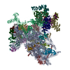
| Title | Structure of mammalian eIF3 in the context of the 43S preinitiation complex | |||||||||
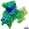
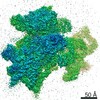
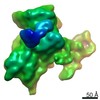
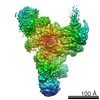
 Map data Map data | Reconstruction of mammalian 43S preinitiation complex bound to DHX29, after focused classification on eIF3 peripheral subunit D. The map was filtered to 10A based on the local resolution of eIF3D | |||||||||
 Sample Sample |
| |||||||||
 Keywords Keywords | eIF3 / eIF3 octamer core / mammalian preinitiation 34S complex / eIF3g/i/b / eIF3d | |||||||||
| Biological species |  | |||||||||
| Method | single particle reconstruction / cryo EM / Resolution: 9.0 Å | |||||||||
 Authors Authors | des-Georges A / Dhote V / Kuhn L / Hellen CUT / Pestova TV / Frank J / Hashem Y | |||||||||
 Citation Citation | Journal: Nature / Year: 2015 Title: Structure of mammalian eIF3 in the context of the 43S preinitiation complex. Authors: Amedee des Georges / Vidya Dhote / Lauriane Kuhn / Christopher U T Hellen / Tatyana V Pestova / Joachim Frank / Yaser Hashem /   Abstract: During eukaryotic translation initiation, 43S complexes, comprising a 40S ribosomal subunit, initiator transfer RNA and initiation factors (eIF) 2, 3, 1 and 1A, attach to the 5'-terminal region of ...During eukaryotic translation initiation, 43S complexes, comprising a 40S ribosomal subunit, initiator transfer RNA and initiation factors (eIF) 2, 3, 1 and 1A, attach to the 5'-terminal region of messenger RNA and scan along it to the initiation codon. Scanning on structured mRNAs also requires the DExH-box protein DHX29. Mammalian eIF3 contains 13 subunits and participates in nearly all steps of translation initiation. Eight subunits having PCI (proteasome, COP9 signalosome, eIF3) or MPN (Mpr1, Pad1, amino-terminal) domains constitute the structural core of eIF3, to which five peripheral subunits are flexibly linked. Here we present a cryo-electron microscopy structure of eIF3 in the context of the DHX29-bound 43S complex, showing the PCI/MPN core at ∼6 Å resolution. It reveals the organization of the individual subunits and their interactions with components of the 43S complex. We were able to build near-complete polyalanine-level models of the eIF3 PCI/MPN core and of two peripheral subunits. The implications for understanding mRNA ribosomal attachment and scanning are discussed. | |||||||||
| History |
|
- Structure visualization
| Movie |
 Movie viewer Movie viewer |
|---|---|
| Structure viewer | EM map: SurfViewMolmilJmol/JSmol |
| Supplemental images |

- Downloads & links
-EMDB archive
| Map data | emd_3058.map.gz | 61.5 MB | EMDB map data format | |
|---|---|---|---|---|
| Header (meta data) | emd-3058-v30.xmlemd-3058.xml | 12.7 KB 12.7 KB | Display Display | EMDB header |
| Images | 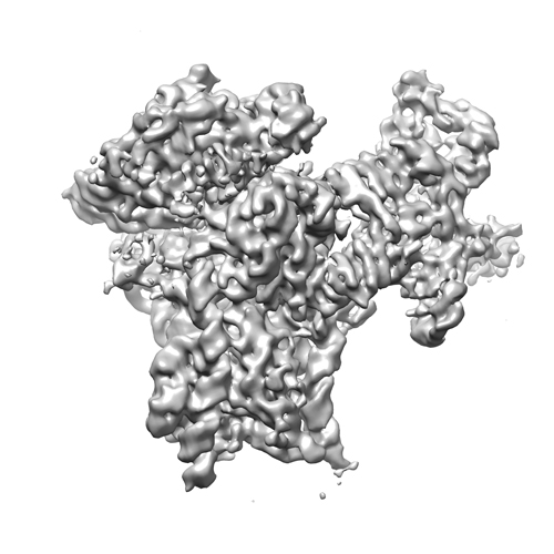 EMD-3058_image.jpg EMD-3058_image.jpg | 112.3 KB | ||
| Archive directory |  http://ftp.pdbj.org/pub/emdb/structures/EMD-3058ftp://ftp.pdbj.org/pub/emdb/structures/EMD-3058 http://ftp.pdbj.org/pub/emdb/structures/EMD-3058ftp://ftp.pdbj.org/pub/emdb/structures/EMD-3058 | HTTPS FTP |
-Related structure data


-Links
| EMDB pages | EMDB (EBI/PDBe) / EMDataResource |
|---|---|
| Related items in Molecule of the Month |

-Map
| File | Download / File: emd_3058.map.gz / Format: CCP4 / Size: 100.6 MB / Type: IMAGE STORED AS FLOATING POINT NUMBER (4 BYTES) | ||||||||||||||||||||||||||||||||||||||||||||||||||||||||||||||||||||
|---|---|---|---|---|---|---|---|---|---|---|---|---|---|---|---|---|---|---|---|---|---|---|---|---|---|---|---|---|---|---|---|---|---|---|---|---|---|---|---|---|---|---|---|---|---|---|---|---|---|---|---|---|---|---|---|---|---|---|---|---|---|---|---|---|---|---|---|---|---|
| Annotation | Reconstruction of mammalian 43S preinitiation complex bound to DHX29, after focused classification on eIF3 peripheral subunit D. The map was filtered to 10A based on the local resolution of eIF3D | ||||||||||||||||||||||||||||||||||||||||||||||||||||||||||||||||||||
| Projections & slices | Image control
Images are generated by Spider. | ||||||||||||||||||||||||||||||||||||||||||||||||||||||||||||||||||||
| Voxel size | X=Y=Z: 1.66 Å | ||||||||||||||||||||||||||||||||||||||||||||||||||||||||||||||||||||
| Density |
| ||||||||||||||||||||||||||||||||||||||||||||||||||||||||||||||||||||
| Symmetry | Space group: 1 | ||||||||||||||||||||||||||||||||||||||||||||||||||||||||||||||||||||
| Details | EMDB XML:
CCP4 map header:
| ||||||||||||||||||||||||||||||||||||||||||||||||||||||||||||||||||||
 Z (Sec.)
 Y (Row.)
 X (Col.)


-Supplemental data
- Sample components
-Entire : Mammalian 43S preinitiation complex bound to DHX29
| Entire | Name: Mammalian 43S preinitiation complex bound to DHX29 |
|---|---|
| Components |
|
-Supramolecule #1000: Mammalian 43S preinitiation complex bound to DHX29
| Supramolecule | Name: Mammalian 43S preinitiation complex bound to DHX29 / type: sample / ID: 1000 / Number unique components: 7 |
|---|
-Supramolecule #1: 40S small ribosomal subunit
| Supramolecule | Name: 40S small ribosomal subunit / type: complex / ID: 1 / Name.synonym: SSU / Recombinant expression: No / Ribosome-details: ribosome-eukaryote: SSU 40S |
|---|---|
| Source (natural) | Organism: |
-Macromolecule #1: eukaryotic initiation factor 3
| Macromolecule | Name: eukaryotic initiation factor 3 / type: protein_or_peptide / ID: 1 / Name.synonym: eIF3 / Oligomeric state: Monomer / Recombinant expression: No |
|---|---|
| Source (natural) | Organism: |
-Macromolecule #2: eukaryotic initiation factor 2
| Macromolecule | Name: eukaryotic initiation factor 2 / type: protein_or_peptide / ID: 2 / Name.synonym: eIF2 / Oligomeric state: Monomer / Recombinant expression: No |
|---|---|
| Source (natural) | Organism: |
-Macromolecule #3: DHX29
| Macromolecule | Name: DHX29 / type: protein_or_peptide / ID: 3 / Oligomeric state: Monomer / Recombinant expression: Yes |
|---|---|
| Source (natural) | Organism: |
| Recombinant expression | Organism:  |
-Macromolecule #4: eIF1
| Macromolecule | Name: eIF1 / type: protein_or_peptide / ID: 4 / Oligomeric state: Monomer / Recombinant expression: Yes |
|---|---|
| Source (natural) | Organism: |
| Recombinant expression | Organism: |
-Macromolecule #5: eIF1A
| Macromolecule | Name: eIF1A / type: protein_or_peptide / ID: 5 / Oligomeric state: Monomer / Recombinant expression: Yes |
|---|---|
| Source (natural) | Organism: |
| Recombinant expression | Organism: |
-Macromolecule #6: initiator transfer RNA
| Macromolecule | Name: initiator transfer RNA / type: rna / ID: 6 / Name.synonym: tRNAiMet / Classification: TRANSFER / Structure: DOUBLE HELIX / Synthetic?: No |
|---|---|
| Source (natural) | Organism: |
-Experimental details
-Structure determination
| Method | cryo EM |
|---|---|
 Processing Processing | single particle reconstruction |
| Aggregation state | particle |
-Sample preparation
| Vitrification | Cryogen name: ETHANE / Chamber humidity: 100 % / Chamber temperature: 120 K / Instrument: FEI VITROBOT MARK IV |
|---|
- Electron microscopy
| Microscope | FEI POLARA 300 |
|---|---|
| Temperature | Min: 80 K / Max: 105 K / Average: 100 K |
| Date | Nov 1, 2013 |
| Image recording | Category: CCD / Film or detector model: GATAN K2 (4k x 4k) / Digitization - Sampling interval: 6.35 µm / Average electron dose: 25 e/Å2 |
| Electron beam | Acceleration voltage: 300 kV / Electron source:  FIELD EMISSION GUN FIELD EMISSION GUN |
| Electron optics | Calibrated magnification: 30120 / Illumination mode: FLOOD BEAM / Imaging mode: BRIGHT FIELD / Cs: 2 mm / Nominal defocus max: 4.5 µm / Nominal defocus min: 0.5 µm |
| Sample stage | Specimen holder model: GATAN HELIUM |
| Experimental equipment |  Model: Tecnai Polara / Image courtesy: FEI Company |
-Image processing
| CTF correction | Details: Each particle |
|---|---|
| Final reconstruction | Applied symmetry - Point group: C1 (asymmetric) / Algorithm: OTHER / Resolution.type: BY AUTHOR / Resolution: 9.0 Å / Resolution method: OTHER / Software - Name: Relion / Number images used: 10062 |
