 Movie
Movie Controller

[English] 日本語
 Yorodumi
- PDB-6w2s: Structure of the Cricket Paralysis Virus 5-UTR IRES (CrPV 5-UTR-I... -
+ Open data

- Basic information
| Entry | Database: PDB / ID: 6w2s | ||||||
|---|---|---|---|---|---|---|---|
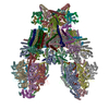
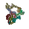
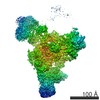
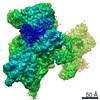
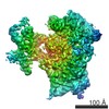
| Title | Structure of the Cricket Paralysis Virus 5-UTR IRES (CrPV 5-UTR-IRES) bound to the small ribosomal subunit in the open state (Class 1) | ||||||
 Components Components |
| ||||||
 Keywords Keywords | Ribosome/TRANSLATION / CrPV 5'-UTR IRES / Internal ribosome entry site / RIBOSOME / Ribosome-TRANSLATION complex | ||||||
| Function / homology |  Function and homology information Function and homology informationviral translational termination-reinitiation / eukaryotic translation initiation factor 3 complex, eIF3e / eukaryotic translation initiation factor 3 complex, eIF3m / IRES-dependent viral translational initiation / translation reinitiation / formation of cytoplasmic translation initiation complex / multi-eIF complex / eukaryotic translation initiation factor 3 complex / eukaryotic 43S preinitiation complex / eukaryotic 48S preinitiation complex ...viral translational termination-reinitiation / eukaryotic translation initiation factor 3 complex, eIF3e / eukaryotic translation initiation factor 3 complex, eIF3m / IRES-dependent viral translational initiation / translation reinitiation / formation of cytoplasmic translation initiation complex / multi-eIF complex / eukaryotic translation initiation factor 3 complex / eukaryotic 43S preinitiation complex / eukaryotic 48S preinitiation complex / regulation of translational initiation / nuclear-transcribed mRNA catabolic process, nonsense-mediated decay / metal-dependent deubiquitinase activity / laminin receptor activity / 90S preribosome / ubiquitin ligase inhibitor activity / positive regulation of signal transduction by p53 class mediator / phagocytic cup / translation regulator activity / rough endoplasmic reticulum / ribosomal small subunit export from nucleus / laminin binding / translation initiation factor binding / translation initiation factor activity / gastrulation / MDM2/MDM4 family protein binding / class I DNA-(apurinic or apyrimidinic site) endonuclease activity / cytosolic ribosome / DNA-(apurinic or apyrimidinic site) lyase / positive regulation of translation / maturation of SSU-rRNA from tricistronic rRNA transcript (SSU-rRNA, 5.8S rRNA, LSU-rRNA) / positive regulation of apoptotic signaling pathway / maturation of SSU-rRNA / small-subunit processome / PML body / spindle / fibrillar center / metallopeptidase activity / rRNA processing / rhythmic process / regulation of translation / positive regulation of canonical Wnt signaling pathway / ribosomal small subunit assembly / virus receptor activity / ribosome binding / ribosomal small subunit biogenesis / small ribosomal subunit / small ribosomal subunit rRNA binding / cytosolic small ribosomal subunit / perikaryon / cell differentiation / cytoplasmic translation / cysteine-type deubiquitinase activity / mitochondrial inner membrane / postsynaptic density / rRNA binding / structural constituent of ribosome / translation / ribonucleoprotein complex / cell division / DNA repair / mRNA binding / apoptotic process / centrosome / synapse / dendrite / nucleolus / perinuclear region of cytoplasm / Golgi apparatus / DNA binding / RNA binding / zinc ion binding / nucleoplasm / membrane / identical protein binding / nucleus / plasma membrane / cytosol Similarity search - Function | ||||||
| Biological species |  Cricket paralysis virus Cricket paralysis virus | ||||||
| Method | ELECTRON MICROSCOPY / single particle reconstruction / cryo EM / Resolution: 3.47 Å | ||||||
 Authors Authors | Neupane, R. / Pisareva, V. / Rodriguez, C.F. / Pisarev, A. / Fernandez, I.S. | ||||||
| Funding support |  United States, 1items United States, 1items
| ||||||
 Citation Citation | Journal: Elife / Year: 2020 Title: A complex IRES at the 5'-UTR of a viral mRNA assembles a functional 48S complex via an uAUG intermediate. Authors: Ritam Neupane / Vera P Pisareva / Carlos F Rodriguez / Andrey V Pisarev / Israel S Fernández /  Abstract: Taking control of the cellular apparatus for protein production is a requirement for virus progression. To ensure this control, diverse strategies of cellular mimicry and/or ribosome hijacking have ...Taking control of the cellular apparatus for protein production is a requirement for virus progression. To ensure this control, diverse strategies of cellular mimicry and/or ribosome hijacking have evolved. The initiation stage of translation is especially targeted as it involves multiple steps and the engagement of numerous initiation factors. The use of structured RNA sequences, called nternal ibosomal ntry ites (IRES), in viral RNAs is a widespread strategy for the exploitation of eukaryotic initiation. Using a combination of electron cryo-microscopy (cryo-EM) and reconstituted translation initiation assays with native components, we characterized how a novel IRES at the 5'-UTR of a viral RNA assembles a functional initiation complex via an uAUG intermediate. The IRES features a novel extended, multi-domain architecture, that circles the 40S head. The structures and accompanying functional data illustrate the importance of 5'-UTR regions in translation regulation and underline the relevance of the untapped diversity of viral IRESs. | ||||||
| History |
|
- Structure visualization
| Movie |
Movie viewer |
|---|---|
| Structure viewer | Molecule: MolmilJmol/JSmol |
- Downloads & links
-Download
| PDBx/mmCIF format | 6w2s.cif.gz | 2.6 MB | Display | PDBx/mmCIF format |
|---|---|---|---|---|
| PDB format | pdb6w2s.ent.gz | Display | PDB format | |
| PDBx/mmJSON format | 6w2s.json.gz | Tree view | PDBx/mmJSON format | |
| Others |  Other downloads Other downloads |
-Validation report
| Arichive directory | https://data.pdbj.org/pub/pdb/validation_reports/w2/6w2sftp://data.pdbj.org/pub/pdb/validation_reports/w2/6w2s | HTTPS FTP |
|---|
-Related structure data
| Related structure data |  21529MC  6w2tC M: map data used to model this data C: citing same article ( |
|---|---|
| Similar structure data |


-Links
 PDBj
PDBj


- Assembly
| Deposited unit | 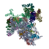
|
|---|---|
| 1 |
|
-Components
-RNA chain , 2 types, 2 molecules A0
| #1: RNA chain | Mass: 547733.062 Da / Num. of mol.: 1 / Source method: isolated from a natural source / Source: (natural) |
|---|---|
| #2: RNA chain | Mass: 121094.953 Da / Num. of mol.: 1 Source method: isolated from a genetically manipulated source Source: (gene. exp.) Cricket paralysis virus / Production host:  |
+Protein , 33 types, 33 molecules BCDFHIJKMOPWXYZbcfEGLNQRSTUVad...
-Eukaryotic translation initiation factor 3 subunit ... , 8 types, 8 molecules 12345678
| #36: Protein | Mass: 164902.656 Da / Num. of mol.: 1 / Source method: isolated from a natural source / Source: (natural) |
|---|---|
| #37: Protein | Mass: 105706.156 Da / Num. of mol.: 1 / Source method: isolated from a natural source / Source: (natural) |
| #38: Protein | Mass: 54199.863 Da / Num. of mol.: 1 / Source method: isolated from a natural source / Source: (natural) |
| #39: Protein | Mass: 37846.730 Da / Num. of mol.: 1 / Source method: isolated from a natural source / Source: (natural) |
| #40: Protein | Mass: 41083.531 Da / Num. of mol.: 1 / Source method: isolated from a natural source / Source: (natural) |
| #41: Protein | Mass: 25129.709 Da / Num. of mol.: 1 / Source method: isolated from a natural source / Source: (natural) |
| #42: Protein | Mass: 71001.477 Da / Num. of mol.: 1 / Source method: isolated from a natural source / Source: (natural) |
| #43: Protein | Mass: 42555.832 Da / Num. of mol.: 1 / Source method: isolated from a natural source / Source: (natural) |
-Non-polymers , 2 types, 2 molecules 

| #44: Chemical | ChemComp-MG /  Mass: 24.305 Da / Num. of mol.: 1 / Source method: obtained synthetically / Formula: Mg Mass: 24.305 Da / Num. of mol.: 1 / Source method: obtained synthetically / Formula: Mg |
|---|---|
| #45: Chemical | ChemComp-ZN /  Mass: 65.409 Da / Num. of mol.: 1 / Source method: obtained synthetically / Formula: Zn Mass: 65.409 Da / Num. of mol.: 1 / Source method: obtained synthetically / Formula: Zn |
-Details
| Has ligand of interest | N |
|---|
-Experimental details
-Experiment
| Experiment | Method: ELECTRON MICROSCOPY |
|---|---|
| EM experiment | Aggregation state: PARTICLE / 3D reconstruction method: single particle reconstruction |
- Sample preparation
| Component | Name: Structure of the Cricket Paralysis Virus 5-UTR IRES (CrPV 5-UTR-IRES) bound to the small ribosomal subunit in the open state (Class 1) Type: RIBOSOME / Entity ID: #1-#43 / Source: MULTIPLE SOURCES |
|---|---|
| Molecular weight | Experimental value: NO |
| Source (natural) | Organism: |
| Buffer solution | pH: 7.5 |
| Specimen | Embedding applied: NO / Shadowing applied: NO / Staining applied: NO / Vitrification applied: YES |
| Specimen support | Grid material: GOLD / Grid mesh size: 300 divisions/in. / Grid type: Quantifoil, UltrAuFoil, R1.2/1.3 |
| Vitrification | Instrument: FEI VITROBOT MARK IV / Cryogen name: ETHANE / Humidity: 100 % / Chamber temperature: 277.15 K Details: Grids were blotted for 2.5s and flash cooled in liquid ethane |
- Electron microscopy imaging
| Experimental equipment |  Model: Titan Krios / Image courtesy: FEI Company |
|---|---|
| Microscopy | Model: FEI TITAN KRIOS |
| Electron gun | Electron source:  FIELD EMISSION GUN / Accelerating voltage: 300 kV / Illumination mode: FLOOD BEAM FIELD EMISSION GUN / Accelerating voltage: 300 kV / Illumination mode: FLOOD BEAM |
| Electron lens | Mode: BRIGHT FIELD |
| Image recording | Average exposure time: 8 sec. / Electron dose: 56.9 e/Å2 / Detector mode: COUNTING / Film or detector model: GATAN K2 SUMMIT (4k x 4k) / Num. of grids imaged: 1 |
| Image scans | Movie frames/image: 40 / Used frames/image: 1-40 |
- Processing
| Software | Name: REFMAC / Version: 5.8.0253 / Classification: refinement | ||||||||||||||||||||||||||||||||||||||||||||||||||||||||||||||||||||||||||||||||||||||||||||||||||||||||||
|---|---|---|---|---|---|---|---|---|---|---|---|---|---|---|---|---|---|---|---|---|---|---|---|---|---|---|---|---|---|---|---|---|---|---|---|---|---|---|---|---|---|---|---|---|---|---|---|---|---|---|---|---|---|---|---|---|---|---|---|---|---|---|---|---|---|---|---|---|---|---|---|---|---|---|---|---|---|---|---|---|---|---|---|---|---|---|---|---|---|---|---|---|---|---|---|---|---|---|---|---|---|---|---|---|---|---|---|
| EM software |
| ||||||||||||||||||||||||||||||||||||||||||||||||||||||||||||||||||||||||||||||||||||||||||||||||||||||||||
| Image processing | Details: The microscope was equipped with an energy filter with slits aperture of 20eV, installed before the detector. | ||||||||||||||||||||||||||||||||||||||||||||||||||||||||||||||||||||||||||||||||||||||||||||||||||||||||||
| CTF correction | Type: PHASE FLIPPING AND AMPLITUDE CORRECTION | ||||||||||||||||||||||||||||||||||||||||||||||||||||||||||||||||||||||||||||||||||||||||||||||||||||||||||
| Particle selection | Num. of particles selected: 915647 | ||||||||||||||||||||||||||||||||||||||||||||||||||||||||||||||||||||||||||||||||||||||||||||||||||||||||||
| Symmetry | Point symmetry: C1 (asymmetric) | ||||||||||||||||||||||||||||||||||||||||||||||||||||||||||||||||||||||||||||||||||||||||||||||||||||||||||
| 3D reconstruction | Resolution: 3.47 Å / Resolution method: FSC 0.143 CUT-OFF / Num. of particles: 36100 / Num. of class averages: 1 / Symmetry type: POINT | ||||||||||||||||||||||||||||||||||||||||||||||||||||||||||||||||||||||||||||||||||||||||||||||||||||||||||
| Refinement | Resolution: 3.47→3.47 Å / Cor.coef. Fo:Fc: 0.843 / SU B: 20.39 / SU ML: 0.323 / ESU R: 0.367 Stereochemistry target values: MAXIMUM LIKELIHOOD WITH PHASES Details: HYDROGENS HAVE BEEN ADDED IN THE RIDING POSITIONS
| ||||||||||||||||||||||||||||||||||||||||||||||||||||||||||||||||||||||||||||||||||||||||||||||||||||||||||
| Solvent computation | Ion probe radii: 0.8 Å / Shrinkage radii: 0.8 Å / VDW probe radii: 1.2 Å / Solvent model: MASK | ||||||||||||||||||||||||||||||||||||||||||||||||||||||||||||||||||||||||||||||||||||||||||||||||||||||||||
| Displacement parameters | Biso mean: 120.284 Å2
| ||||||||||||||||||||||||||||||||||||||||||||||||||||||||||||||||||||||||||||||||||||||||||||||||||||||||||
| Refinement step | Cycle: 1 / Total: 106817 | ||||||||||||||||||||||||||||||||||||||||||||||||||||||||||||||||||||||||||||||||||||||||||||||||||||||||||
| Refine LS restraints |
|