 Movie
Movie Controller

[English] 日本語
 Yorodumi
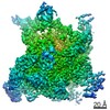
- EMDB-21531: Head masked 3D refined density of the Cricket Paralysis Virus 5-U... -
+ Open data

- Basic information
| Entry | Database: EMDB / ID: EMD-21531 | |||||||||
|---|---|---|---|---|---|---|---|---|---|---|
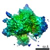
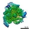
| Title | Head masked 3D refined density of the Cricket Paralysis Virus 5-UTR IRES (CrPV 5-UTR-IRES) bound to the small ribosomal subunit used to build the 5-UTR IRES model | |||||||||
 Map data Map data | Sharpened map after post processing for head masked | |||||||||
 Sample Sample |
| |||||||||
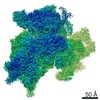
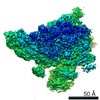
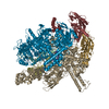
| Biological species |  | |||||||||
| Method | single particle reconstruction / cryo EM / Resolution: 2.79 Å | |||||||||
 Authors Authors | Neupane R / Pisareva V / Rodriguez CF / Pisarev A / Fernandez IS | |||||||||
| Funding support |  United States, 1 items United States, 1 items
| |||||||||
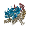
 Citation Citation | Journal: Elife / Year: 2020 Title: A complex IRES at the 5'-UTR of a viral mRNA assembles a functional 48S complex via an uAUG intermediate. Authors: Ritam Neupane / Vera P Pisareva / Carlos F Rodriguez / Andrey V Pisarev / Israel S Fernández /  Abstract: Taking control of the cellular apparatus for protein production is a requirement for virus progression. To ensure this control, diverse strategies of cellular mimicry and/or ribosome hijacking have ...Taking control of the cellular apparatus for protein production is a requirement for virus progression. To ensure this control, diverse strategies of cellular mimicry and/or ribosome hijacking have evolved. The initiation stage of translation is especially targeted as it involves multiple steps and the engagement of numerous initiation factors. The use of structured RNA sequences, called nternal ibosomal ntry ites (IRES), in viral RNAs is a widespread strategy for the exploitation of eukaryotic initiation. Using a combination of electron cryo-microscopy (cryo-EM) and reconstituted translation initiation assays with native components, we characterized how a novel IRES at the 5'-UTR of a viral RNA assembles a functional initiation complex via an uAUG intermediate. The IRES features a novel extended, multi-domain architecture, that circles the 40S head. The structures and accompanying functional data illustrate the importance of 5'-UTR regions in translation regulation and underline the relevance of the untapped diversity of viral IRESs. | |||||||||
| History |
|
- Structure visualization
| Movie |
 Movie viewer Movie viewer |
|---|---|
| Structure viewer | EM map: SurfViewMolmilJmol/JSmol |
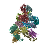
| Supplemental images |

- Downloads & links
-EMDB archive
| Map data | emd_21531.map.gz | 17.4 MB | EMDB map data format | |
|---|---|---|---|---|
| Header (meta data) | emd-21531-v30.xmlemd-21531.xml | 22.2 KB 22.2 KB | Display Display | EMDB header |
| FSC (resolution estimation) | emd_21531_fsc.xml | 14.2 KB | Display | FSC data file |
| Images | 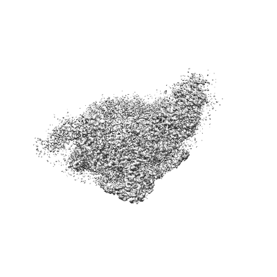 emd_21531.png emd_21531.png | 28.6 KB | ||
| Masks | emd_21531_msk_1.map | 244.1 MB | Mask map | |
| Others | emd_21531_additional.map.gzemd_21531_half_map_1.map.gzemd_21531_half_map_2.map.gz | 193.4 MB 194.1 MB 194.2 MB | ||
| Archive directory |  http://ftp.pdbj.org/pub/emdb/structures/EMD-21531ftp://ftp.pdbj.org/pub/emdb/structures/EMD-21531 http://ftp.pdbj.org/pub/emdb/structures/EMD-21531ftp://ftp.pdbj.org/pub/emdb/structures/EMD-21531 | HTTPS FTP |
-Related structure data


-Links
| EMDB pages | EMDB (EBI/PDBe) / EMDataResource |
|---|---|
| Related items in Molecule of the Month |

-Map
| File | Download / File: emd_21531.map.gz / Format: CCP4 / Size: 244.1 MB / Type: IMAGE STORED AS FLOATING POINT NUMBER (4 BYTES) | ||||||||||||||||||||||||||||||||||||||||||||||||||||||||||||||||||||
|---|---|---|---|---|---|---|---|---|---|---|---|---|---|---|---|---|---|---|---|---|---|---|---|---|---|---|---|---|---|---|---|---|---|---|---|---|---|---|---|---|---|---|---|---|---|---|---|---|---|---|---|---|---|---|---|---|---|---|---|---|---|---|---|---|---|---|---|---|---|
| Annotation | Sharpened map after post processing for head masked | ||||||||||||||||||||||||||||||||||||||||||||||||||||||||||||||||||||
| Projections & slices | Image control
Images are generated by Spider. | ||||||||||||||||||||||||||||||||||||||||||||||||||||||||||||||||||||
| Voxel size | X=Y=Z: 1.0605 Å | ||||||||||||||||||||||||||||||||||||||||||||||||||||||||||||||||||||
| Density |
| ||||||||||||||||||||||||||||||||||||||||||||||||||||||||||||||||||||
| Symmetry | Space group: 1 | ||||||||||||||||||||||||||||||||||||||||||||||||||||||||||||||||||||
| Details | EMDB XML:
CCP4 map header:
| ||||||||||||||||||||||||||||||||||||||||||||||||||||||||||||||||||||
 Z (Sec.)
 Y (Row.)
 X (Col.)


-Supplemental data
-Mask #1
| File | emd_21531_msk_1.map | ||||||||||||
|---|---|---|---|---|---|---|---|---|---|---|---|---|---|
| Projections & Slices |
| ||||||||||||
| Density Histograms |


-Additional map: Unsharpened map before post-processing for head masked
| File | emd_21531_additional.map | ||||||||||||
|---|---|---|---|---|---|---|---|---|---|---|---|---|---|
| Annotation | Unsharpened map before post-processing for head masked | ||||||||||||
| Projections & Slices |
| ||||||||||||
| Density Histograms |


-Half map: Half map 1 for head masked
| File | emd_21531_half_map_1.map | ||||||||||||
|---|---|---|---|---|---|---|---|---|---|---|---|---|---|
| Annotation | Half map 1 for head masked | ||||||||||||
| Projections & Slices |
| ||||||||||||
| Density Histograms |


-Half map: Half map 2 for head masked
| File | emd_21531_half_map_2.map | ||||||||||||
|---|---|---|---|---|---|---|---|---|---|---|---|---|---|
| Annotation | Half map 2 for head masked | ||||||||||||
| Projections & Slices |
| ||||||||||||
| Density Histograms |


- Sample components
-Entire : Cricket Paralysis Virus 5-UTR IRES (CrPV 5-UTR-IRES) bound to the...
| Entire | Name: Cricket Paralysis Virus 5-UTR IRES (CrPV 5-UTR-IRES) bound to the small ribosomal subunit from a head masked 3D refinement |
|---|---|
| Components |
|
-Supramolecule #1: Cricket Paralysis Virus 5-UTR IRES (CrPV 5-UTR-IRES) bound to the...
| Supramolecule | Name: Cricket Paralysis Virus 5-UTR IRES (CrPV 5-UTR-IRES) bound to the small ribosomal subunit from a head masked 3D refinement type: complex / ID: 1 / Parent: 0 / Macromolecule list: #1-#43 |
|---|---|
| Source (natural) | Organism: |
-Experimental details
-Structure determination
| Method | cryo EM |
|---|---|
 Processing Processing | single particle reconstruction |
| Aggregation state | particle |
-Sample preparation
| Buffer | pH: 7.5 |
|---|---|
| Vitrification | Cryogen name: ETHANE / Chamber humidity: 100 % / Chamber temperature: 277.15 K / Instrument: FEI VITROBOT MARK IV Details: Grids were blotted for 2.5s and flash cooled in liquid ethane. |
- Electron microscopy
| Microscope | FEI TITAN KRIOS |
|---|---|
| Image recording | Film or detector model: GATAN K2 SUMMIT (4k x 4k) / Detector mode: COUNTING / Digitization - Frames/image: 1-40 / Number grids imaged: 1 / Average exposure time: 8.0 sec. / Average electron dose: 56.9 e/Å2 |
| Electron beam | Acceleration voltage: 300 kV / Electron source:  FIELD EMISSION GUN FIELD EMISSION GUN |
| Electron optics | Illumination mode: FLOOD BEAM / Imaging mode: BRIGHT FIELD |
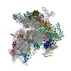
| Experimental equipment |  Model: Titan Krios / Image courtesy: FEI Company |

