- EMDB-21530: Structure of the Cricket Paralysis Virus 5-UTR IRES (CrPV 5-UTR-I... -
+
Open data
ID or keywords:
Loading...
-
Basic information
Entry
Database: EMDB / ID: EMD-21530
Title
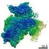
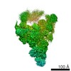
Structure of the Cricket Paralysis Virus 5-UTR IRES (CrPV 5-UTR-IRES) bound to the small ribosomal subunit in the closed state (Class 2)
Map data
Sharpened map after post processing for Class 2 (closed)
Sample
Complex: Structure of the Cricket Paralysis Virus 5-UTR IRES (CrPV 5-UTR-IRES) bound to the small ribosomal subunit in the closed state (Class 2)
RNA: x 2 types
Protein or peptide: x 42 types
Ligand: x 2 types
Keywords
CrPV 5'-UTR IRES / Internal ribosome entry site / RIBOSOME
Function / homology
Function and homology information
viral translational termination-reinitiation / eukaryotic translation initiation factor 3 complex, eIF3e / cap-dependent translational initiation / eukaryotic translation initiation factor 3 complex, eIF3m / IRES-dependent viral translational initiation / translation reinitiation / formation of cytoplasmic translation initiation complex / multi-eIF complex / eukaryotic translation initiation factor 3 complex / eukaryotic 43S preinitiation complex ...viral translational termination-reinitiation / eukaryotic translation initiation factor 3 complex, eIF3e / cap-dependent translational initiation / eukaryotic translation initiation factor 3 complex, eIF3m / IRES-dependent viral translational initiation / translation reinitiation / formation of cytoplasmic translation initiation complex / multi-eIF complex / eukaryotic translation initiation factor 3 complex / eukaryotic 43S preinitiation complex / mRNA cap binding / eukaryotic 48S preinitiation complex / regulation of translational initiation / nuclear-transcribed mRNA catabolic process, nonsense-mediated decay / metal-dependent deubiquitinase activity / laminin receptor activity / ubiquitin ligase inhibitor activity / 90S preribosome / positive regulation of signal transduction by p53 class mediator / phagocytic cup / translation regulator activity / rough endoplasmic reticulum / laminin binding / ribosomal small subunit export from nucleus / translation initiation factor activity / gastrulation / translation initiation factor binding / MDM2/MDM4 family protein binding / class I DNA-(apurinic or apyrimidinic site) endonuclease activity / cytosolic ribosome / DNA-(apurinic or apyrimidinic site) lyase / positive regulation of translation / maturation of SSU-rRNA from tricistronic rRNA transcript (SSU-rRNA, 5.8S rRNA, LSU-rRNA) / positive regulation of apoptotic signaling pathway / maturation of SSU-rRNA / small-subunit processome / PML body / spindle / fibrillar center / metallopeptidase activity / rRNA processing / rhythmic process / positive regulation of canonical Wnt signaling pathway / regulation of translation / ribosomal small subunit assembly / virus receptor activity / ribosome binding / ribosomal small subunit biogenesis / small ribosomal subunit / small ribosomal subunit rRNA binding / cytosolic small ribosomal subunit / perikaryon / cell differentiation / cytoplasmic translation / cysteine-type deubiquitinase activity / mitochondrial inner membrane / postsynaptic density / rRNA binding / structural constituent of ribosome / translation / ribonucleoprotein complex / cell division / DNA repair / mRNA binding / apoptotic process / centrosome / synapse / dendrite / nucleolus / perinuclear region of cytoplasm / Golgi apparatus / DNA binding / RNA binding / zinc ion binding / nucleoplasm / membrane / identical protein binding / nucleus / plasma membrane / cytosol Similarity search - Function
Eukaryotic translation initiation factor 3 subunit D / Eukaryotic translation initiation factor 3 subunit 7 (eIF-3) / Eukaryotic translation initiation factor 3 subunit H / eIF3h, C-terminal / C-terminal region of eIF3h / Eukaryotic translation initiation factor 3 subunit F / Translation initiation factor 3 complex subunit L / RNA polymerase I-associated factor PAF67 / Eukaryotic translation initiation factor 3 subunit M / eIF3 subunit M, C-terminal helix domain ...Eukaryotic translation initiation factor 3 subunit D / Eukaryotic translation initiation factor 3 subunit 7 (eIF-3) / Eukaryotic translation initiation factor 3 subunit H / eIF3h, C-terminal / C-terminal region of eIF3h / Eukaryotic translation initiation factor 3 subunit F / Translation initiation factor 3 complex subunit L / RNA polymerase I-associated factor PAF67 / Eukaryotic translation initiation factor 3 subunit M / eIF3 subunit M, C-terminal helix domain / : / eIF3 subunit 6 N terminal domain / eIF3 subunit M, C-terminal helix / Eukaryotic translation initiation factor 3 subunit E, C-terminal / EIF3CL-like, C-terminal domain / Eukaryotic translation initiation factor 3 subunit E / Eukaryotic translation initiation factor 3 subunit E, N-terminal / eIF3 subunit 6 N terminal domain / Eukaryotic translation initiation factor 3 subunit K / Translation initiation factor 3, subunit 12, N-terminal, eukaryotic / : / eIF3a, PCI domain, TPR-like region / Eukaryotic translation initiation factor 3 subunit A / Eukaryotic translation initiation factor 3 subunit C, N-terminal domain / Eukaryotic translation initiation factor 3 subunit C / Eukaryotic translation initiation factor 3 subunit 8 N-terminus / Eukaryotic translation initiation factor 3 subunit M eIF3m/COP9 signalosome complex subunit 7 COPS7 / CSN8/PSMD8/EIF3K / CSN8/PSMD8/EIF3K family / Rpn11/EIF3F, C-terminal / Maintenance of mitochondrial structure and function / motif in proteasome subunits, Int-6, Nip-1 and TRIP-15 / : / PCI domain / Proteasome component (PCI) domain / PCI domain profile. / 40S ribosomal protein SA / 40S ribosomal protein SA, C-terminal domain / 40S ribosomal protein SA C-terminus / Ubiquitin-like protein FUBI / JAB1/Mov34/MPN/PAD-1 ubiquitin protease / : / Ribosomal protein S26e signature. / : / Ribosomal protein S12e signature. / Ribosomal protein S26e / Ribosomal protein S26e superfamily / Ribosomal protein S26e / Ribosomal protein S12e / Small (40S) ribosomal subunit Asc1/RACK1 / Ribosomal protein S5, eukaryotic/archaeal / Ribosomal protein S19e, conserved site / Ribosomal protein S19e signature. / Ribosomal protein S2, eukaryotic / 40S Ribosomal protein S10 / S27a-like superfamily / JAB/MPN domain / Plectin/S10, N-terminal / Plectin/S10 domain / JAB1/MPN/MOV34 metalloenzyme domain / Ribosomal protein S10, eukaryotic/archaeal / Ribosomal protein S30 / Ribosomal protein S30 / Ribosomal protein S25 / S25 ribosomal protein / Ribosomal protein S8e subdomain, eukaryotes / : / Ribosomal protein S17e, conserved site / Ribosomal protein S17e signature. / Ribosomal protein S7e signature. / Ribosomal protein S27a / Ribosomal protein S27a / Ribosomal protein S27a / Ribosomal protein S2, eukaryotic/archaeal / MPN domain / MPN domain profile. / 40S ribosomal protein S29/30S ribosomal protein S14 type Z / Ribosomal protein S3, eukaryotic/archaeal / Ribosomal protein S3Ae, conserved site / Ribosomal protein S3Ae signature. / Ribosomal protein S8e, conserved site / Ribosomal protein S8e signature. / Ribosomal protein S27e signature. / 40S ribosomal protein S4, C-terminal domain / 40S ribosomal protein S4 C-terminus / Ribosomal protein S19A/S15e / Ribosomal protein S19e / Ribosomal protein S19e / Ribosomal_S19e / Ribosomal protein S4e, N-terminal, conserved site / Ribosomal protein S4e signature. / Ribosomal protein S17e / Ribosomal protein S17e-like superfamily / Ribosomal S17 / Ribosomal protein S6, eukaryotic / 40S ribosomal protein S1/3, eukaryotes / 40S ribosomal protein S11, N-terminal / Ribosomal_S17 N-terminal / Ribosomal protein S7e / Ribosomal protein S7e Similarity search - Domain/homology
Small ribosomal subunit protein uS4 / Eukaryotic translation initiation factor 3 subunit L / Small ribosomal subunit protein eS12 / Small ribosomal subunit protein uS9 / Small ribosomal subunit protein uS10 / Small ribosomal subunit protein RACK1 / Ubiquitin-ribosomal protein eS31 fusion protein / Eukaryotic translation initiation factor 3 subunit F / Eukaryotic translation initiation factor 3 subunit M / Eukaryotic translation initiation factor 3 subunit A ...Small ribosomal subunit protein uS4 / Eukaryotic translation initiation factor 3 subunit L / Small ribosomal subunit protein eS12 / Small ribosomal subunit protein uS9 / Small ribosomal subunit protein uS10 / Small ribosomal subunit protein RACK1 / Ubiquitin-ribosomal protein eS31 fusion protein / Eukaryotic translation initiation factor 3 subunit F / Eukaryotic translation initiation factor 3 subunit M / Eukaryotic translation initiation factor 3 subunit A / Small ribosomal subunit protein uS15 / Small ribosomal subunit protein eS1 / Eukaryotic translation initiation factor 3 subunit H / Eukaryotic translation initiation factor 3 subunit E / Small ribosomal subunit protein eS7 / Small ribosomal subunit protein uS5 / Small ribosomal subunit protein uS12 / 40S ribosomal protein S24 / Eukaryotic translation initiation factor 3 subunit K / Ubiquitin-like FUBI-ribosomal protein eS30 fusion protein / Small ribosomal subunit protein eS25 / Small ribosomal subunit protein eS26 / Small ribosomal subunit protein uS7 / Small ribosomal subunit protein uS8 / Small ribosomal subunit protein eS28 / Small ribosomal subunit protein eS8 / Small ribosomal subunit protein eS4 / Small ribosomal subunit protein eS6 / Small ribosomal subunit protein eS19 / Small ribosomal subunit protein uS3 / Small ribosomal subunit protein uS13 / Small ribosomal subunit protein eS10 / Small ribosomal subunit protein uS17 / Small ribosomal subunit protein eS17 / Small ribosomal subunit protein uS2 / Small ribosomal subunit protein eS27 / Small ribosomal subunit protein uS19 / Small ribosomal subunit protein uS11 / Small ribosomal subunit protein uS14 / Eukaryotic translation initiation factor 3 subunit C / Eukaryotic translation initiation factor 3 subunit D Similarity search - Component
National Institutes of Health/National Institute of General Medical Sciences (NIH/NIGMS)
GM097014
United States
Citation
Journal: Elife / Year: 2020 Title: A complex IRES at the 5'-UTR of a viral mRNA assembles a functional 48S complex via an uAUG intermediate. Authors: Ritam Neupane / Vera P Pisareva / Carlos F Rodriguez / Andrey V Pisarev / Israel S Fernández / Abstract: Taking control of the cellular apparatus for protein production is a requirement for virus progression. To ensure this control, diverse strategies of cellular mimicry and/or ribosome hijacking have ...Taking control of the cellular apparatus for protein production is a requirement for virus progression. To ensure this control, diverse strategies of cellular mimicry and/or ribosome hijacking have evolved. The initiation stage of translation is especially targeted as it involves multiple steps and the engagement of numerous initiation factors. The use of structured RNA sequences, called nternal ibosomal ntry ites (IRES), in viral RNAs is a widespread strategy for the exploitation of eukaryotic initiation. Using a combination of electron cryo-microscopy (cryo-EM) and reconstituted translation initiation assays with native components, we characterized how a novel IRES at the 5'-UTR of a viral RNA assembles a functional initiation complex via an uAUG intermediate. The IRES features a novel extended, multi-domain architecture, that circles the 40S head. The structures and accompanying functional data illustrate the importance of 5'-UTR regions in translation regulation and underline the relevance of the untapped diversity of viral IRESs.
History
Deposition
Mar 8, 2020
-
Header (metadata) release
Apr 22, 2020
-
Map release
Apr 22, 2020
-
Update
Mar 20, 2024
-
Current status
Mar 20, 2024
Processing site: RCSB / Status: Released
-
Structure visualization
Movie
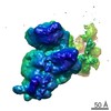
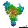
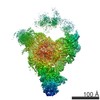
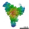
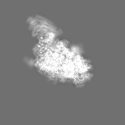
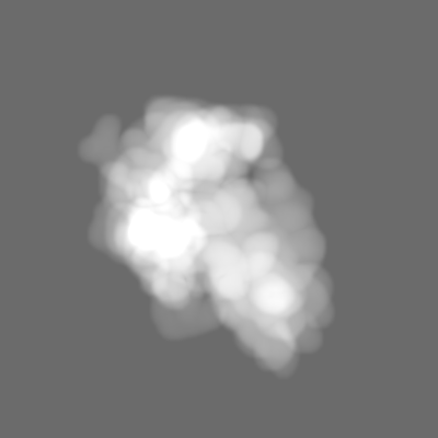
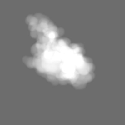
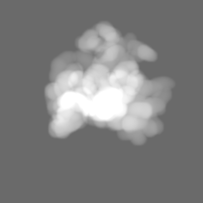
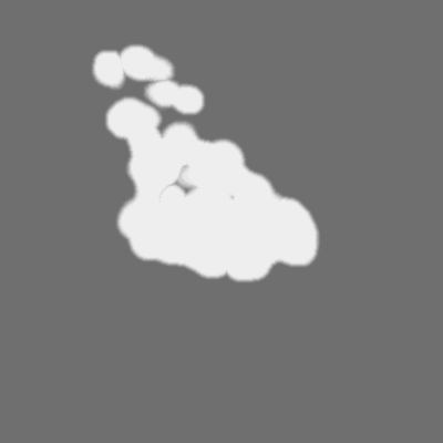
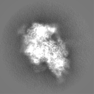
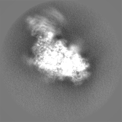
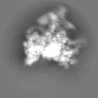
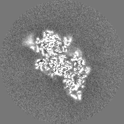
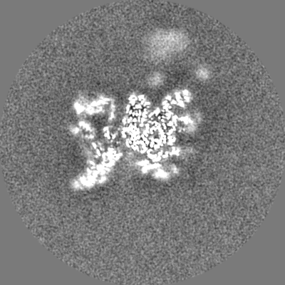
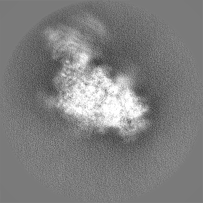
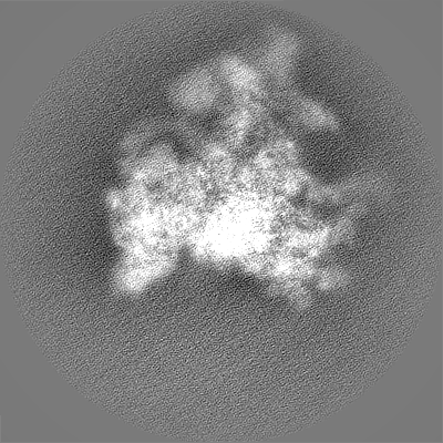
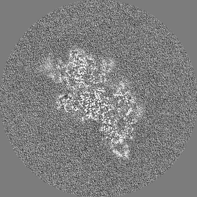
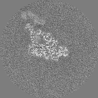
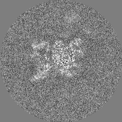
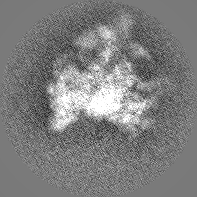
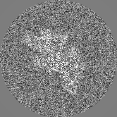
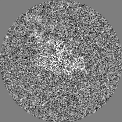
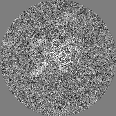
Surface view with section colored by density value
Entire : Structure of the Cricket Paralysis Virus 5-UTR IRES (CrPV 5-UTR-I...
Entire
Name: Structure of the Cricket Paralysis Virus 5-UTR IRES (CrPV 5-UTR-IRES) bound to the small ribosomal subunit in the closed state (Class 2)
Components
Complex: Structure of the Cricket Paralysis Virus 5-UTR IRES (CrPV 5-UTR-IRES) bound to the small ribosomal subunit in the closed state (Class 2)
RNA: 18S rRNA
Protein or peptide: uS2
Protein or peptide: eS1
Protein or peptide: uS5
Protein or peptide: eS4
Protein or peptide: eS6
Protein or peptide: eS7
Protein or peptide: eS8
Protein or peptide: uS4
Protein or peptide: uS17
Protein or peptide: uS15
Protein or peptide: uS11
Protein or peptide: eS21
Protein or peptide: uS8
Protein or peptide: uS12
Protein or peptide: eS24
Protein or peptide: eS26
Protein or peptide: eS27
Protein or peptide: eS30
Protein or peptide: uS3
Protein or peptide: uS7
Protein or peptide: eS10
Protein or peptide: eS12
Protein or peptide: uS19
Protein or peptide: uS9
Protein or peptide: eS17
Protein or peptide: uS13
Protein or peptide: eS19
Protein or peptide: uS10
Protein or peptide: eS25
Protein or peptide: eS28
Protein or peptide: eS29
Protein or peptide: eS31
Protein or peptide: RACK1
Protein or peptide: Eukaryotic translation initiation factor 3 subunit E
Protein or peptide: Eukaryotic translation initiation factor 3 subunit F
Protein or peptide: Eukaryotic translation initiation factor 3 subunit K
Protein or peptide: Eukaryotic translation initiation factor 3 subunit L
Protein or peptide: Eukaryotic translation initiation factor 3 subunit M
Protein or peptide: Eukaryotic translation initiation factor 3 subunit A
Protein or peptide: Eukaryotic translation initiation factor 3 subunit C
Protein or peptide: Eukaryotic translation initiation factor 3 subunit D
Protein or peptide: Eukaryotic translation initiation factor 3 subunit H
RNA: CrPV 5'-UTR IRES
Ligand: MAGNESIUM ION
Ligand: ZINC ION
+
Supramolecule #1: Structure of the Cricket Paralysis Virus 5-UTR IRES (CrPV 5-UTR-I...
Supramolecule
Name: Structure of the Cricket Paralysis Virus 5-UTR IRES (CrPV 5-UTR-IRES) bound to the small ribosomal subunit in the closed state (Class 2) type: complex / ID: 1 / Parent: 0 / Macromolecule list: #1-#44
Source (natural)
Organism: Oryctolagus cuniculus (rabbit)
+
Macromolecule #1: 18S rRNA
Macromolecule
Name: 18S rRNA / type: rna / ID: 1 / Number of copies: 1
Cryogen name: ETHANE / Chamber humidity: 100 % / Chamber temperature: 277.15 K / Instrument: FEI VITROBOT MARK IV Details: Grids were blotted for 2.5s and flash cooled in liquid ethane.
-
Electron microscopy
Microscope
FEI TITAN KRIOS
Image recording
Film or detector model: GATAN K2 SUMMIT (4k x 4k) / Detector mode: COUNTING / Digitization - Frames/image: 1-40 / Number grids imaged: 1 / Average exposure time: 8.0 sec. / Average electron dose: 56.9 e/Å2
Electron beam
Acceleration voltage: 300 kV / Electron source: FIELD EMISSION GUN
Electron optics
Illumination mode: FLOOD BEAM / Imaging mode: BRIGHT FIELD
Experimental equipment
Model: Titan Krios / Image courtesy: FEI Company
+
Image processing
Details
The microscope was equipped with an energy filter with slits aperture of 20eV, installed before the detector.
Particle selection
Number selected: 915647
Startup model
Type of model: EMDB MAP
Final reconstruction
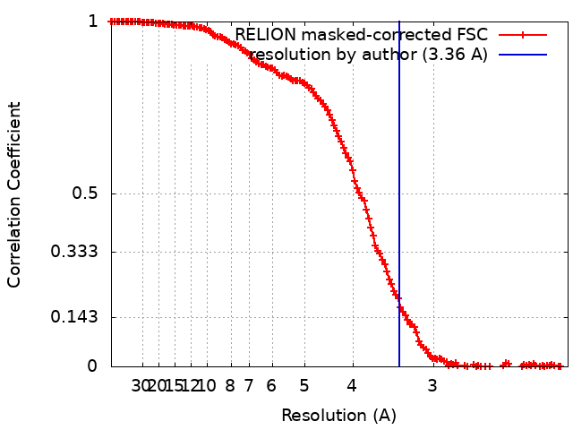
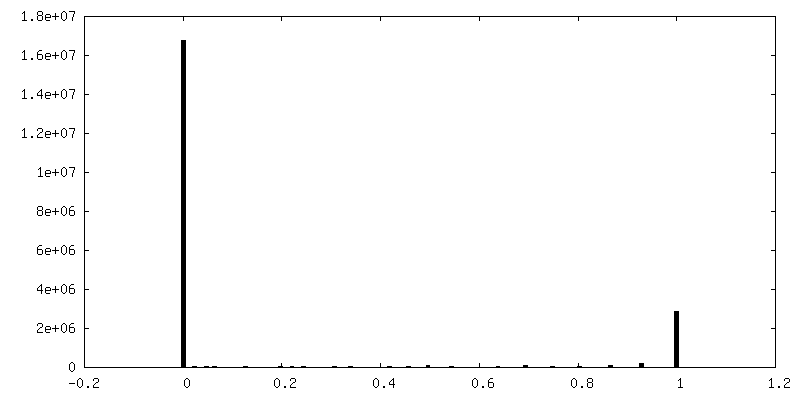
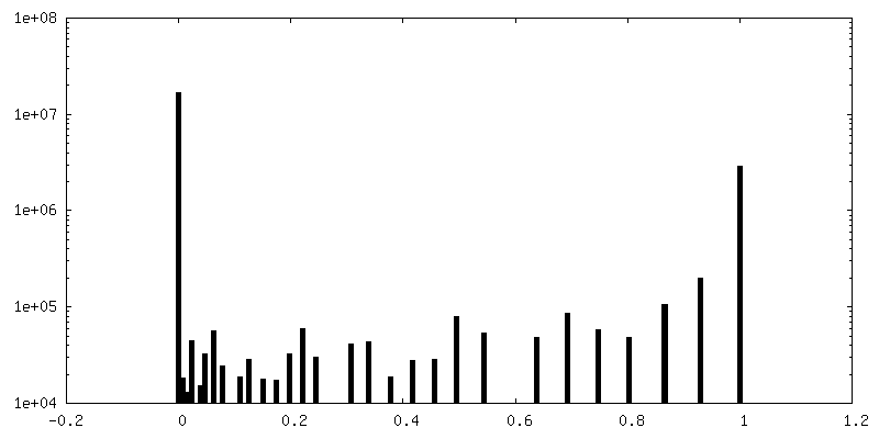
Number classes used: 1 / Applied symmetry - Point group: C1 (asymmetric) / Resolution.type: BY AUTHOR / Resolution: 3.36 Å / Resolution method: FSC 0.143 CUT-OFF / Software - Name: RELION / Number images used: 23444
Initial angle assignment
Type: MAXIMUM LIKELIHOOD / Software - Name: RELION
Final angle assignment
Type: MAXIMUM LIKELIHOOD / Software - Name: RELION
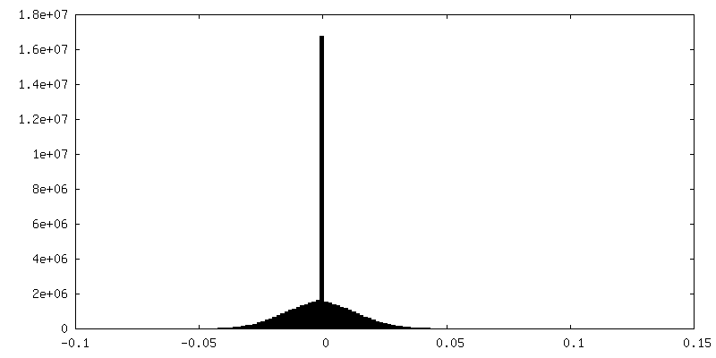
FSC plot (resolution estimation)
+
About Yorodumi
-
News
-
Feb 9, 2022. New format data for meta-information of EMDB entries
New format data for meta-information of EMDB entries
Version 3 of the EMDB header file is now the official format.
The previous official version 1.9 will be removed from the archive.
In the structure databanks used in Yorodumi, some data are registered as the other names, "COVID-19 virus" and "2019-nCoV". Here are the details of the virus and the list of structure data.
Jan 31, 2019. EMDB accession codes are about to change! (news from PDBe EMDB page)
EMDB accession codes are about to change! (news from PDBe EMDB page)
The allocation of 4 digits for EMDB accession codes will soon come to an end. Whilst these codes will remain in use, new EMDB accession codes will include an additional digit and will expand incrementally as the available range of codes is exhausted. The current 4-digit format prefixed with “EMD-” (i.e. EMD-XXXX) will advance to a 5-digit format (i.e. EMD-XXXXX), and so on. It is currently estimated that the 4-digit codes will be depleted around Spring 2019, at which point the 5-digit format will come into force.
The EM Navigator/Yorodumi systems omit the EMD- prefix.
Related info.:Q: What is EMD? / ID/Accession-code notation in Yorodumi/EM Navigator
Yorodumi is a browser for structure data from EMDB, PDB, SASBDB, etc.
This page is also the successor to EM Navigator detail page, and also detail information page/front-end page for Omokage search.
The word "yorodu" (or yorozu) is an old Japanese word meaning "ten thousand". "mi" (miru) is to see.
Related info.:EMDB / PDB / SASBDB / Comparison of 3 databanks / Yorodumi Search / Aug 31, 2016. New EM Navigator & Yorodumi / Yorodumi Papers / Jmol/JSmol / Function and homology information / Changes in new EM Navigator and Yorodumi
 Movie
Movie Controller

 Yorodumi
 Open data
 Basic information
 Map data
 Sample
 Keywords
 Function and homology information
 Cricket paralysis virus
 Authors
 United States, 1 items
  Citation
 Structure visualization
 Downloads & links
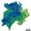
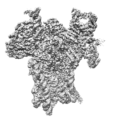 emd_21530.png
 http://ftp.pdbj.org/pub/emdb/structures/EMD-21530
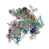


 Z (Sec.)
 Y (Row.)
 X (Col.)


 Sample components
 Processing
 Electron microscopy
 FIELD EMISSION GUN