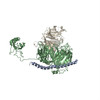
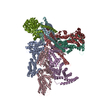
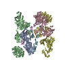
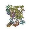
Journal: Nature / Year: 2015 Title: Structure of mammalian eIF3 in the context of the 43S preinitiation complex. Authors: Amedee des Georges / Vidya Dhote / Lauriane Kuhn / Christopher U T Hellen / Tatyana V Pestova / Joachim Frank / Yaser Hashem / Abstract: During eukaryotic translation initiation, 43S complexes, comprising a 40S ribosomal subunit, initiator transfer RNA and initiation factors (eIF) 2, 3, 1 and 1A, attach to the 5'-terminal region of ...During eukaryotic translation initiation, 43S complexes, comprising a 40S ribosomal subunit, initiator transfer RNA and initiation factors (eIF) 2, 3, 1 and 1A, attach to the 5'-terminal region of messenger RNA and scan along it to the initiation codon. Scanning on structured mRNAs also requires the DExH-box protein DHX29. Mammalian eIF3 contains 13 subunits and participates in nearly all steps of translation initiation. Eight subunits having PCI (proteasome, COP9 signalosome, eIF3) or MPN (Mpr1, Pad1, amino-terminal) domains constitute the structural core of eIF3, to which five peripheral subunits are flexibly linked. Here we present a cryo-electron microscopy structure of eIF3 in the context of the DHX29-bound 43S complex, showing the PCI/MPN core at ∼6 Å resolution. It reveals the organization of the individual subunits and their interactions with components of the 43S complex. We were able to build near-complete polyalanine-level models of the eIF3 PCI/MPN core and of two peripheral subunits. The implications for understanding mRNA ribosomal attachment and scanning are discussed.
History
Deposition
Jun 21, 2015
-
Header (metadata) release
Jul 8, 2015
-
Map release
Sep 9, 2015
-
Update
Oct 7, 2015
-
Current status
Oct 7, 2015
Processing site: PDBe / Status: Released
-
Structure visualization
Movie
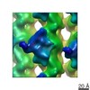
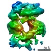
Surface view with section colored by density value
Protein or peptide: eukaryotic initiation factor 3
Protein or peptide: eukaryotic initiation factor 2
Protein or peptide: DHX29
Protein or peptide: eukaryotic initiation factor 1
Protein or peptide: eukaryotic initiation factor 1A
RNA: initiator transfer RNA
-
Supramolecule #1000: eIF3 octamer core of Rabbit eIF3
Supramolecule
Name: eIF3 octamer core of Rabbit eIF3 / type: sample / ID: 1000 Details: The sample was monodisperse, eIF3 octamer core was locally refined from a reconstruction of the mammalian 43S preinitiation complex Oligomeric state: One octamer / Number unique components: 7
Cryogen name: ETHANE / Chamber humidity: 100 % / Chamber temperature: 120 K / Instrument: FEI VITROBOT MARK IV
-
Electron microscopy
Microscope
FEI POLARA 300
Temperature
Min: 80 K / Max: 105 K / Average: 100 K
Date
Nov 1, 2013
Image recording
Category: CCD / Film or detector model: GATAN K2 (4k x 4k) / Digitization - Sampling interval: 6.35 µm / Number real images: 8000 / Average electron dose: 25 e/Å2
Tilt angle min
0
Tilt angle max
0
Electron beam
Acceleration voltage: 300 kV / Electron source: FIELD EMISSION GUN
In the structure databanks used in Yorodumi, some data are registered as the other names, "COVID-19 virus" and "2019-nCoV". Here are the details of the virus and the list of structure data.
Jan 31, 2019. EMDB accession codes are about to change! (news from PDBe EMDB page)
EMDB accession codes are about to change! (news from PDBe EMDB page)
The allocation of 4 digits for EMDB accession codes will soon come to an end. Whilst these codes will remain in use, new EMDB accession codes will include an additional digit and will expand incrementally as the available range of codes is exhausted. The current 4-digit format prefixed with “EMD-” (i.e. EMD-XXXX) will advance to a 5-digit format (i.e. EMD-XXXXX), and so on. It is currently estimated that the 4-digit codes will be depleted around Spring 2019, at which point the 5-digit format will come into force.
The EM Navigator/Yorodumi systems omit the EMD- prefix.
Related info.:Q: What is EMD? / ID/Accession-code notation in Yorodumi/EM Navigator
Yorodumi is a browser for structure data from EMDB, PDB, SASBDB, etc.
This page is also the successor to EM Navigator detail page, and also detail information page/front-end page for Omokage search.
The word "yorodu" (or yorozu) is an old Japanese word meaning "ten thousand". "mi" (miru) is to see.
Related info.:EMDB / PDB / SASBDB / Comparison of 3 databanks / Yorodumi Search / Aug 31, 2016. New EM Navigator & Yorodumi / Yorodumi Papers / Jmol/JSmol / Function and homology information / Changes in new EM Navigator and Yorodumi
 Movie
Movie Controller

 Yorodumi
 Open data
 Basic information
 Map data
 Sample
 Keywords
 Function and homology information
 Authors
 Citation

 Structure visualization
 Downloads & links
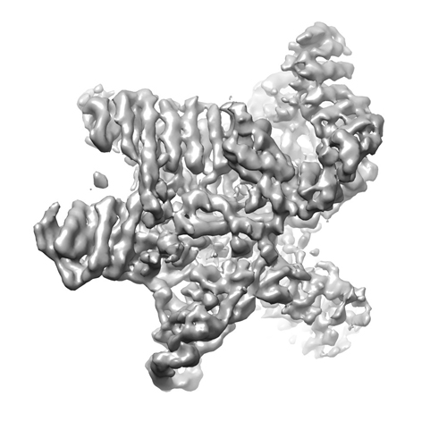 13501.jpg
 http://ftp.pdbj.org/pub/emdb/structures/EMD-3056


 Z (Sec.)
 Y (Row.)
 X (Col.)


 Sample components
 Processing
 Electron microscopy
 FIELD EMISSION GUN
