 Movie
Movie Controller

[English] 日本語
 Yorodumi
- PDB-1ryz: Uridine Phosphorylase from Salmonella typhimurium. Crystal Struct... -
+ Open data

- Basic information
| Entry | Database: PDB / ID: 1ryz | ||||||
|---|---|---|---|---|---|---|---|
| Title | Uridine Phosphorylase from Salmonella typhimurium. Crystal Structure at 2.9 A Resolution | ||||||
 Components Components | Uridine phosphorylase | ||||||
 Keywords Keywords | TRANSFERASE / uridine phosphorylase / nucleoside phosphorylase | ||||||
| Function / homology |  Function and homology information Function and homology informationnucleotide catabolic process / uridine phosphorylase / nucleoside catabolic process / UMP salvage / uridine phosphorylase activity / cytosol Similarity search - Function | ||||||
| Biological species |  Salmonella typhimurium (bacteria) Salmonella typhimurium (bacteria) | ||||||
| Method |  X-RAY DIFFRACTION / MOLECULAR REPLACEMENT / Resolution: 2.9 Å X-RAY DIFFRACTION / MOLECULAR REPLACEMENT / Resolution: 2.9 Å | ||||||
 Authors Authors | Dontsova, M.V. / Gabdoulkhakov, A.G. / Lyashenko, A.V. / Nikonov, S.V. / Ealick, S.E. / Mikhailov, A.M. | ||||||
 Citation Citation | Journal: TO BE PUBLISHED Title: Structure-functions studies of uridine phosphorylase from Salmonella typhimurium Authors: Dontsova, M.V. / Gabdoulkhakov, A.G. / Lyashenko, A.V. / Nikonov, S.V. / Ealick, S.E. / Mikhailov, A.M. | ||||||
| History |
|
- Structure visualization
| Structure viewer | Molecule: MolmilJmol/JSmol |
|---|
- Downloads & links
-Download
| PDBx/mmCIF format | 1ryz.cif.gz | 276.2 KB | Display | PDBx/mmCIF format |
|---|---|---|---|---|
| PDB format | pdb1ryz.ent.gz | 223.4 KB | Display | PDB format |
| PDBx/mmJSON format | 1ryz.json.gz | Tree view | PDBx/mmJSON format | |
| Others |  Other downloads Other downloads |
-Validation report
| Arichive directory | https://data.pdbj.org/pub/pdb/validation_reports/ry/1ryzftp://data.pdbj.org/pub/pdb/validation_reports/ry/1ryz | HTTPS FTP |
|---|
-Related structure data
| Related structure data |  1k3fS S: Starting model for refinement |
|---|---|
| Similar structure data |

-Links
 PDBj
PDBj

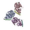
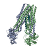
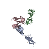
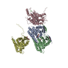
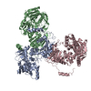
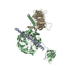
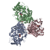
- Assembly
| Deposited unit | 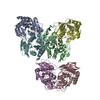
| ||||||||
|---|---|---|---|---|---|---|---|---|---|
| 1 |
| ||||||||
| 2 | 
| ||||||||
| 3 | 
| ||||||||
| 4 | 
| ||||||||
| Unit cell |
|
-Components
| #1: Protein | Mass: 27169.092 Da / Num. of mol.: 6 Source method: isolated from a genetically manipulated source Source: (gene. exp.) Salmonella typhimurium (bacteria) / Strain: LT2 / Gene: udp / Plasmid: pBluescript IISK / Species (production host): Escherichia coli / Production host: #2: Chemical |   Mass: 60.052 Da / Num. of mol.: 2 / Source method: obtained synthetically / Formula: C2H4O2 Mass: 60.052 Da / Num. of mol.: 2 / Source method: obtained synthetically / Formula: C2H4O2 |
|---|
-Experimental details
-Experiment
| Experiment | Method: X-RAY DIFFRACTION / Number of used crystals: 1 |
|---|
- Sample preparation
| Crystal | Density Matthews: 2.02 Å3/Da / Density % sol: 38.98 % |
|---|---|
| Crystal grow | Temperature: 294 K / Method: vapor diffusion, hanging drop / pH: 4.6 Details: 0.1M Na Acetate trihydrate, 10% PEG 8000, pH 4.6, VAPOR DIFFUSION, HANGING DROP, temperature 294K |
-Data collection
| Diffraction | Mean temperature: 110 K |
|---|---|
| Diffraction source | Source: ROTATING ANODE / Type: OTHER / Wavelength: 1.54179 Å |
| Detector | Type: MARRESEARCH / Detector: IMAGE PLATE / Date: Aug 15, 2003 / Details: Capillary optics |
| Radiation | Monochromator: GRAPHITE / Protocol: SINGLE WAVELENGTH / Monochromatic (M) / Laue (L): M / Scattering type: x-ray |
| Radiation wavelength | Wavelength: 1.54179 Å / Relative weight: 1 |
| Reflection | Resolution: 2.9→30 Å / Num. all: 75053 / Num. obs: 26462 / % possible obs: 93 % / Observed criterion σ(F): 0 / Observed criterion σ(I): 0 / Redundancy: 2.84 % / Biso Wilson estimate: -0.2 Å2 / Rmerge(I) obs: 0.154 / Rsym value: 0.177 / Net I/σ(I): 6.89 |
| Reflection shell | Resolution: 2.9→2.95 Å / Redundancy: 2.79 % / Rmerge(I) obs: 0.338 / Mean I/σ(I) obs: 3.05 / Num. unique all: 1074 / Rsym value: 0.333 / % possible all: 75.9 |
- Processing
| Software |
| ||||||||||||||||||||||||||||||||||||
|---|---|---|---|---|---|---|---|---|---|---|---|---|---|---|---|---|---|---|---|---|---|---|---|---|---|---|---|---|---|---|---|---|---|---|---|---|---|
| Refinement | Method to determine structure: MOLECULAR REPLACEMENT Starting model: PDB entry 1K3F Resolution: 2.9→29.46 Å / Rfactor Rfree error: 0.008 / Data cutoff high absF: 3363606.7 / Data cutoff low absF: 0 / Isotropic thermal model: RESTRAINED / Cross valid method: THROUGHOUT / σ(F): 2 / Stereochemistry target values: Engh & Huber / Details: BULK SOLVENT MODEL USED
| ||||||||||||||||||||||||||||||||||||
| Solvent computation | Solvent model: FLAT MODEL / Bsol: 44.0393 Å2 / ksol: 0.301264 e/Å3 | ||||||||||||||||||||||||||||||||||||
| Displacement parameters | Biso mean: 18.5 Å2
| ||||||||||||||||||||||||||||||||||||
| Refine analyze |
| ||||||||||||||||||||||||||||||||||||
| Refinement step | Cycle: LAST / Resolution: 2.9→29.46 Å
| ||||||||||||||||||||||||||||||||||||
| Refine LS restraints |
| ||||||||||||||||||||||||||||||||||||
| LS refinement shell | Resolution: 2.9→3.08 Å / Rfactor Rfree error: 0.02 / Total num. of bins used: 6
| ||||||||||||||||||||||||||||||||||||
| Xplor file |
|
