 Movie
Movie Controller

[English] 日本語
 Yorodumi
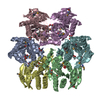
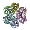
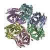
- PDB-1k3f: Uridine Phosphorylase from E. coli, Refined in the Monoclinic Cry... -
+ Open data

- Basic information
| Entry | Database: PDB / ID: 1k3f | ||||||
|---|---|---|---|---|---|---|---|
| Title | Uridine Phosphorylase from E. coli, Refined in the Monoclinic Crystal Lattice | ||||||
 Components Components | uridine phosphorylase | ||||||
 Keywords Keywords | TRANSFERASE / nucleoside phosphorylase / hexamer | ||||||
| Function / homology |  Function and homology information Function and homology informationUMP catabolic process / uridine catabolic process / uridine phosphorylase / UMP salvage / uridine phosphorylase activity / potassium ion binding / DNA damage response / protein-containing complex / ATP binding / identical protein binding / cytosol Similarity search - Function | ||||||
| Biological species |  | ||||||
| Method |  X-RAY DIFFRACTION / MOLECULAR REPLACEMENT / Resolution: 2.5 Å X-RAY DIFFRACTION / MOLECULAR REPLACEMENT / Resolution: 2.5 Å | ||||||
 Authors Authors | Morgunova, E.Yu. / Mikhailov, A.M. / Popov, A.N. / Blagova, E.V. / Smirnova, E.A. / Vainshtein, B.K. / Mao, C. / Armstrong, S.R. / Ealick, S.E. / Komissarov, A.A. ...Morgunova, E.Yu. / Mikhailov, A.M. / Popov, A.N. / Blagova, E.V. / Smirnova, E.A. / Vainshtein, B.K. / Mao, C. / Armstrong, S.R. / Ealick, S.E. / Komissarov, A.A. / Linkova, E.V. / Burlakova, A.A. / Mironov, A.S. / Debabov, V.G. | ||||||
 Citation Citation | Journal: FEBS Lett. / Year: 1995 Title: Atomic structure at 2.5 A resolution of uridine phosphorylase from E. coli as refined in the monoclinic crystal lattice. Authors: Morgunova, E.Yu. / Mikhailov, A.M. / Popov, A.N. / Blagova, E.V. / Smirnova, E.A. / Vainshtein, B.K. / Mao, C. / Armstrong, S.R. / Ealick, S.E. / Komissarov, A.A. / Linkova, E.V. / ...Authors: Morgunova, E.Yu. / Mikhailov, A.M. / Popov, A.N. / Blagova, E.V. / Smirnova, E.A. / Vainshtein, B.K. / Mao, C. / Armstrong, S.R. / Ealick, S.E. / Komissarov, A.A. / Linkova, E.V. / Burlakova, A.A. / Mironov, A.S. / Debabov, V.G. | ||||||
| History |
|
- Structure visualization
| Structure viewer | Molecule: MolmilJmol/JSmol |
|---|
- Downloads & links
-Download
| PDBx/mmCIF format | 1k3f.cif.gz | 283.4 KB | Display | PDBx/mmCIF format |
|---|---|---|---|---|
| PDB format | pdb1k3f.ent.gz | 226.1 KB | Display | PDB format |
| PDBx/mmJSON format | 1k3f.json.gz | Tree view | PDBx/mmJSON format | |
| Others |  Other downloads Other downloads |
-Validation report
| Arichive directory | https://data.pdbj.org/pub/pdb/validation_reports/k3/1k3fftp://data.pdbj.org/pub/pdb/validation_reports/k3/1k3f | HTTPS FTP |
|---|
-Related structure data
| Similar structure data |
|---|


-Links
 PDBj
PDBj- Assembly
| Deposited unit | 
| ||||||||
|---|---|---|---|---|---|---|---|---|---|
| 1 |
| ||||||||
| Unit cell |
| ||||||||
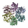
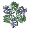
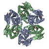
| Details | The biological assembly is a hexamer of identical subunits; asymmetric unit contains a Upase hexamer. Entire hexamer is deposited, |
-Components
| #1: Protein | Mass: 27189.055 Da / Num. of mol.: 6 Source method: isolated from a genetically manipulated source Source: (gene. exp.) |
|---|
-Experimental details
-Experiment
| Experiment | Method: X-RAY DIFFRACTION / Number of used crystals: 1 |
|---|
- Sample preparation
| Crystal | Density Matthews: 2.24 Å3/Da / Density % sol: 47.1 % | |||||||||||||||||||||||||||||||||||
|---|---|---|---|---|---|---|---|---|---|---|---|---|---|---|---|---|---|---|---|---|---|---|---|---|---|---|---|---|---|---|---|---|---|---|---|---|
| Crystal grow | Temperature: 293 K / Method: vapor diffusion, sitting drop / pH: 7.3 Details: drops-0.05 M Tris-HCl and 4-6% PEG 4000; equilibrium solution-0.1M Tris-mal/NaOH pH5.91-5.96, 20-25% Peg 4000 and 0.04% sodium azide, pH 7.3, VAPOR DIFFUSION, SITTING DROP, temperature 293K | |||||||||||||||||||||||||||||||||||
| Crystal grow | *PLUS | |||||||||||||||||||||||||||||||||||
| Components of the solutions | *PLUS
|
-Data collection
| Diffraction | Mean temperature: 293 K |
|---|---|
| Diffraction source | Source: ROTATING ANODE / Type: OTHER / Wavelength: 1.5418 |
| Detector | Type: KARD-6 / Detector: DIFFRACTOMETER / Date: Jan 5, 1994 |
| Radiation | Monochromator: graphite / Protocol: SINGLE WAVELENGTH / Monochromatic (M) / Laue (L): M / Scattering type: x-ray |
| Radiation wavelength | Wavelength: 1.5418 Å / Relative weight: 1 |
| Reflection | Resolution: 2.5→40 Å / Num. all: 50479 / Num. obs: 42403 / % possible obs: 84 % / Observed criterion σ(F): 0 / Observed criterion σ(I): 2 / Rmerge(I) obs: 0.097 |
| Reflection | *PLUS Lowest resolution: 40 Å / % possible obs: 84 % / Num. measured all: 116050 |
- Processing
| Software |
| ||||||||||||||||||||
|---|---|---|---|---|---|---|---|---|---|---|---|---|---|---|---|---|---|---|---|---|---|
| Refinement | Method to determine structure: MOLECULAR REPLACEMENT Starting model: trigonal form of Upase Resolution: 2.5→6 Å / Isotropic thermal model: isotropic / σ(F): 2 / Stereochemistry target values: Engh & Huber
| ||||||||||||||||||||
| Refinement step | Cycle: LAST / Resolution: 2.5→6 Å
| ||||||||||||||||||||
| Refine LS restraints |
| ||||||||||||||||||||
| Software | *PLUS Name: X-PLOR / Version: 3.1 / Classification: refinement | ||||||||||||||||||||
| Refinement | *PLUS Highest resolution: 2.5 Å / Lowest resolution: 6 Å / σ(F): 2 | ||||||||||||||||||||
| Solvent computation | *PLUS | ||||||||||||||||||||
| Displacement parameters | *PLUS | ||||||||||||||||||||
| Refine LS restraints | *PLUS Type: x_bond_d / Dev ideal: 0.012 |
