 Movie
Movie Controller

[English] 日本語
 Yorodumi
- PDB-5cws: Crystal structure of the intact Chaetomium thermophilum Nsp1-Nup4... -
+ Open data

- Basic information
| Entry | Database: PDB / ID: 5cws | ||||||||||||||||||
|---|---|---|---|---|---|---|---|---|---|---|---|---|---|---|---|---|---|---|---|
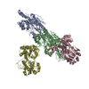
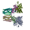
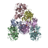
| Title | Crystal structure of the intact Chaetomium thermophilum Nsp1-Nup49-Nup57 channel nucleoporin heterotrimer bound to its Nic96 nuclear pore complex attachment site | ||||||||||||||||||
 Components Components |
| ||||||||||||||||||
 Keywords Keywords | PROTEIN TRANSPORT / nucleocytoplasmic transport | ||||||||||||||||||
| Function / homology |  Function and homology information Function and homology informationnuclear pore central transport channel / protein localization to nuclear inner membrane / nuclear pore organization / structural constituent of nuclear pore / RNA export from nucleus / nuclear localization sequence binding / NLS-bearing protein import into nucleus / poly(A)+ mRNA export from nucleus / nuclear pore / mRNA transport ...nuclear pore central transport channel / protein localization to nuclear inner membrane / nuclear pore organization / structural constituent of nuclear pore / RNA export from nucleus / nuclear localization sequence binding / NLS-bearing protein import into nucleus / poly(A)+ mRNA export from nucleus / nuclear pore / mRNA transport / phospholipid binding / protein import into nucleus / protein transport / nuclear membrane Similarity search - Function | ||||||||||||||||||
| Biological species |  Homo sapiens (human) Homo sapiens (human) Chaetomium thermophilum (fungus) Chaetomium thermophilum (fungus) | ||||||||||||||||||
| Method |  X-RAY DIFFRACTION / SYNCHROTRON / AB INITIO PHASING / Resolution: 3.77 Å X-RAY DIFFRACTION / SYNCHROTRON / AB INITIO PHASING / Resolution: 3.77 Å | ||||||||||||||||||
 Authors Authors | Bley, C.J. / Petrovic, S. / Paduch, M. / Lu, V. / Kossiakoff, A.A. / Hoelz, A. | ||||||||||||||||||
| Funding support |  United States, 5items United States, 5items
| ||||||||||||||||||
 Citation Citation | Journal: Science / Year: 2015 Title: Architecture of the fungal nuclear pore inner ring complex. Authors: Stuwe, T. / Bley, C.J. / Thierbach, K. / Petrovic, S. / Schilbach, S. / Mayo, D.J. / Perriches, T. / Rundlet, E.J. / Jeon, Y.E. / Collins, L.N. / Huber, F.M. / Lin, D.H. / Paduch, M. / ...Authors: Stuwe, T. / Bley, C.J. / Thierbach, K. / Petrovic, S. / Schilbach, S. / Mayo, D.J. / Perriches, T. / Rundlet, E.J. / Jeon, Y.E. / Collins, L.N. / Huber, F.M. / Lin, D.H. / Paduch, M. / Koide, A. / Lu, V. / Fischer, J. / Hurt, E. / Koide, S. / Kossiakoff, A.A. / Hoelz, A. | ||||||||||||||||||
| History |
|
- Structure visualization
| Structure viewer | Molecule: MolmilJmol/JSmol |
|---|
- Downloads & links
-Download
| PDBx/mmCIF format | 5cws.cif.gz | 787.1 KB | Display | PDBx/mmCIF format |
|---|---|---|---|---|
| PDB format | pdb5cws.ent.gz | 655 KB | Display | PDB format |
| PDBx/mmJSON format | 5cws.json.gz | Tree view | PDBx/mmJSON format | |
| Others |  Other downloads Other downloads |
-Validation report
| Arichive directory | https://data.pdbj.org/pub/pdb/validation_reports/cw/5cwsftp://data.pdbj.org/pub/pdb/validation_reports/cw/5cws | HTTPS FTP |
|---|
-Related structure data
| Related structure data |  4jnuC  4jnvC  4jo7C  4jo9C  4jq5C 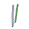 5cwtC  5cwuC  5cwvC  5cwwC C: citing same article ( |
|---|---|
| Similar structure data |


-Links
 PDBj
PDBj


- Assembly
| Deposited unit | 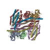
| ||||||||
|---|---|---|---|---|---|---|---|---|---|
| 1 | 
| ||||||||
| 2 | 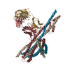
| ||||||||
| Unit cell |
|
-Components
-Protein , 4 types, 8 molecules CIDJEKFL
| #3: Protein | Mass: 23558.211 Da / Num. of mol.: 2 / Fragment: UNP residues 467-674 Source method: isolated from a genetically manipulated source Source: (gene. exp.) Chaetomium thermophilum (strain DSM 1495 / CBS 144.50 / IMI 039719) (fungus)Strain: DSM 1495 / CBS 144.50 / IMI 039719 / Gene: NSP1, CTHT_0054390 / Production host:  #4: Protein | Mass: 24536.504 Da / Num. of mol.: 2 / Fragment: UNP residues 246-470 Source method: isolated from a genetically manipulated source Source: (gene. exp.) Chaetomium thermophilum (fungus) / Strain: DSM 1495 / CBS 144.50 / IMI 039719 / Gene: NUP49, CTHT_0031980 / Production host: #5: Protein | Mass: 28792.684 Da / Num. of mol.: 2 / Fragment: UNP residues 74-319 Source method: isolated from a genetically manipulated source Source: (gene. exp.) Chaetomium thermophilum (strain DSM 1495 / CBS 144.50 / IMI 039719) (fungus)Strain: DSM 1495 / CBS 144.50 / IMI 039719 / Gene: NUP57, CTHT_0010940 / Production host: #6: Protein | Mass: 7793.831 Da / Num. of mol.: 2 / Fragment: UNP residues 139-211 Source method: isolated from a genetically manipulated source Source: (gene. exp.) Chaetomium thermophilum (strain DSM 1495 / CBS 144.50 / IMI 039719) (fungus)Strain: DSM 1495 / CBS 144.50 / IMI 039719 / Gene: NIC96, CTHT_0008480 / Production host: |
|---|
-Antibody , 2 types, 4 molecules AGBH
| #1: Antibody | Mass: 25394.451 Da / Num. of mol.: 2 Source method: isolated from a genetically manipulated source Source: (gene. exp.) Homo sapiens (human) / Production host: #2: Antibody | Mass: 28606.086 Da / Num. of mol.: 2 Source method: isolated from a genetically manipulated source Source: (gene. exp.) Homo sapiens (human) / Production host: |
|---|
-Non-polymers , 1 types, 2 molecules 
| #7: Chemical |  Mass: 190.230 Da / Num. of mol.: 2 / Source method: obtained synthetically / Formula: Os Mass: 190.230 Da / Num. of mol.: 2 / Source method: obtained synthetically / Formula: Os |
|---|
-Details
| Has protein modification | Y |
|---|
-Experimental details
-Experiment
| Experiment | Method: X-RAY DIFFRACTION / Number of used crystals: 1 |
|---|
- Sample preparation
| Crystal | Density Matthews: 3.85 Å3/Da / Density % sol: 68.07 % |
|---|---|
| Crystal grow | Temperature: 294 K / Method: vapor diffusion, hanging drop / pH: 8.7 Details: 0.1M TRIS, pH 8.7 5.7 % (w/v) PEG 20,000 1 % (v/v) 1-propanol PH range: 8.7 - 8.9 |
-Data collection
| Diffraction | Mean temperature: 100 K |
|---|---|
| Diffraction source | Source: SYNCHROTRON / Site: SSRL / Beamline: BL12-2 / Wavelength: 1.14 Å |
| Detector | Type: DECTRIS PILATUS 6M / Detector: PIXEL / Date: Mar 25, 2015 |
| Radiation | Protocol: SINGLE WAVELENGTH / Monochromatic (M) / Laue (L): M / Scattering type: x-ray |
| Radiation wavelength | Wavelength: 1.14 Å / Relative weight: 1 |
| Reflection | Resolution: 3.77→50 Å / Num. obs: 43896 / % possible obs: 99.9 % / Redundancy: 40.2 % / Rmerge(I) obs: 0.204 / Net I/σ(I): 17 |
| Reflection shell | Highest resolution: 3.77 Å |
- Processing
| Software |
| ||||||||||||||||||||||||||||||||||||||||||||||||||||||||||||||||||||||||||||||||||||||||||||||||||||||||||||||||||||||||||||||||||||||||||||||||||||||||||||||||||||||||||||||||||||||||||||||||||||
|---|---|---|---|---|---|---|---|---|---|---|---|---|---|---|---|---|---|---|---|---|---|---|---|---|---|---|---|---|---|---|---|---|---|---|---|---|---|---|---|---|---|---|---|---|---|---|---|---|---|---|---|---|---|---|---|---|---|---|---|---|---|---|---|---|---|---|---|---|---|---|---|---|---|---|---|---|---|---|---|---|---|---|---|---|---|---|---|---|---|---|---|---|---|---|---|---|---|---|---|---|---|---|---|---|---|---|---|---|---|---|---|---|---|---|---|---|---|---|---|---|---|---|---|---|---|---|---|---|---|---|---|---|---|---|---|---|---|---|---|---|---|---|---|---|---|---|---|---|---|---|---|---|---|---|---|---|---|---|---|---|---|---|---|---|---|---|---|---|---|---|---|---|---|---|---|---|---|---|---|---|---|---|---|---|---|---|---|---|---|---|---|---|---|---|---|---|---|
| Refinement | Method to determine structure: AB INITIO PHASING / Resolution: 3.77→49.666 Å / SU ML: 0.64 / Cross valid method: FREE R-VALUE / σ(F): 1.33 / Phase error: 30.79 / Stereochemistry target values: ML
| ||||||||||||||||||||||||||||||||||||||||||||||||||||||||||||||||||||||||||||||||||||||||||||||||||||||||||||||||||||||||||||||||||||||||||||||||||||||||||||||||||||||||||||||||||||||||||||||||||||
| Solvent computation | Shrinkage radii: 0.9 Å / VDW probe radii: 1.11 Å / Solvent model: FLAT BULK SOLVENT MODEL | ||||||||||||||||||||||||||||||||||||||||||||||||||||||||||||||||||||||||||||||||||||||||||||||||||||||||||||||||||||||||||||||||||||||||||||||||||||||||||||||||||||||||||||||||||||||||||||||||||||
| Refinement step | Cycle: LAST / Resolution: 3.77→49.666 Å
| ||||||||||||||||||||||||||||||||||||||||||||||||||||||||||||||||||||||||||||||||||||||||||||||||||||||||||||||||||||||||||||||||||||||||||||||||||||||||||||||||||||||||||||||||||||||||||||||||||||
| Refine LS restraints |
| ||||||||||||||||||||||||||||||||||||||||||||||||||||||||||||||||||||||||||||||||||||||||||||||||||||||||||||||||||||||||||||||||||||||||||||||||||||||||||||||||||||||||||||||||||||||||||||||||||||
| LS refinement shell |
|
