 Movie
Movie Controller


+ Open data

- Basic information
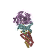
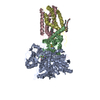
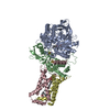
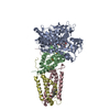
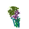
| Entry | Database: PDB / ID: 3cir | ||||||
|---|---|---|---|---|---|---|---|
| Title | E. coli Quinol fumarate reductase FrdA T234A mutation | ||||||
 Components Components | (Fumarate reductase ...) x 4 | ||||||
 Keywords Keywords | OXIDOREDUCTASE / electron transport / Tricarboxylic acid cycle | ||||||
| Function / homology |  Function and homology information Function and homology informationfumarate reductase complex / fermentation / succinate dehydrogenase activity / fumarate metabolic process / anaerobic electron transport chain / succinate dehydrogenase (quinone) activity / succinate dehydrogenase / anaerobic respiration / 3 iron, 4 sulfur cluster binding / bacterial-type flagellum assembly ...fumarate reductase complex / fermentation / succinate dehydrogenase activity / fumarate metabolic process / anaerobic electron transport chain / succinate dehydrogenase (quinone) activity / succinate dehydrogenase / anaerobic respiration / 3 iron, 4 sulfur cluster binding / bacterial-type flagellum assembly / iron-sulfur cluster binding / tricarboxylic acid cycle / FAD binding / 2 iron, 2 sulfur cluster binding / flavin adenine dinucleotide binding / 4 iron, 4 sulfur cluster binding / electron transfer activity / DNA damage response / membrane / metal ion binding / plasma membrane / cytosol Similarity search - Function | ||||||
| Biological species |  | ||||||
| Method |  X-RAY DIFFRACTION / SYNCHROTRON / MOLECULAR REPLACEMENT / Resolution: 3.65 Å X-RAY DIFFRACTION / SYNCHROTRON / MOLECULAR REPLACEMENT / Resolution: 3.65 Å | ||||||
 Authors Authors | Tomasiak, T.M. / Maklashina, E. / Cecchini, G. / Iverson, T.M. | ||||||
 Citation Citation | Journal: J.Biol.Chem. / Year: 2008 Title: A threonine on the active site loop controls transition state formation in Escherichia coli respiratory complex II. Authors: Tomasiak, T.M. / Maklashina, E. / Cecchini, G. / Iverson, T.M. | ||||||
| History |
|
- Structure visualization
| Structure viewer | Molecule: MolmilJmol/JSmol |
|---|
- Downloads & links
-Download
| PDBx/mmCIF format | 3cir.cif.gz | 390.2 KB | Display | PDBx/mmCIF format |
|---|---|---|---|---|
| PDB format | pdb3cir.ent.gz | 313.5 KB | Display | PDB format |
| PDBx/mmJSON format | 3cir.json.gz | Tree view | PDBx/mmJSON format | |
| Others |  Other downloads Other downloads |
-Validation report
| Arichive directory | https://data.pdbj.org/pub/pdb/validation_reports/ci/3cirftp://data.pdbj.org/pub/pdb/validation_reports/ci/3cir | HTTPS FTP |
|---|
-Related structure data
| Related structure data |  2b76S S: Starting model for refinement |
|---|---|
| Similar structure data |


-Links
 PDBj
PDBj


- Assembly
| Deposited unit | 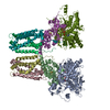
| ||||||||
|---|---|---|---|---|---|---|---|---|---|
| 1 | 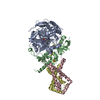
| ||||||||
| 2 | 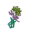
| ||||||||
| Unit cell |
|
-Components
-Fumarate reductase ... , 4 types, 8 molecules AMBNCODP
| #1: Protein | Mass: 66027.523 Da / Num. of mol.: 2 / Mutation: T234A Source method: isolated from a genetically manipulated source Source: (gene. exp.) #2: Protein | Mass: 27021.885 Da / Num. of mol.: 2 Source method: isolated from a genetically manipulated source Source: (gene. exp.) #3: Protein | Mass: 14898.773 Da / Num. of mol.: 2 Source method: isolated from a genetically manipulated source Source: (gene. exp.) #4: Protein | Mass: 13118.870 Da / Num. of mol.: 2 Source method: isolated from a genetically manipulated source Source: (gene. exp.) |
|---|
-Non-polymers , 4 types, 8 molecules 


| #5: Chemical |  Mass: 785.550 Da / Num. of mol.: 2 / Source method: obtained synthetically / Formula: C27H33N9O15P2 / Comment: FAD*YM Mass: 785.550 Da / Num. of mol.: 2 / Source method: obtained synthetically / Formula: C27H33N9O15P2 / Comment: FAD*YM#6: Chemical |  Mass: 175.820 Da / Num. of mol.: 2 / Source method: obtained synthetically / Formula: Fe2S2 Mass: 175.820 Da / Num. of mol.: 2 / Source method: obtained synthetically / Formula: Fe2S2#7: Chemical |  Mass: 295.795 Da / Num. of mol.: 2 / Source method: obtained synthetically / Formula: Fe3S4 Mass: 295.795 Da / Num. of mol.: 2 / Source method: obtained synthetically / Formula: Fe3S4#8: Chemical |  Mass: 351.640 Da / Num. of mol.: 2 / Source method: obtained synthetically / Formula: Fe4S4 Mass: 351.640 Da / Num. of mol.: 2 / Source method: obtained synthetically / Formula: Fe4S4 |
|---|
-Experimental details
-Experiment
| Experiment | Method: X-RAY DIFFRACTION / Number of used crystals: 1 |
|---|
- Sample preparation
| Crystal | Density Matthews: 3.6 Å3/Da / Density % sol: 65.87 % |
|---|---|
| Crystal grow | Temperature: 298 K / Method: vapor diffusion, hanging drop / pH: 5.8 Details: 10% PEG 5000 MME, 0.250M magnesium acetate, 100mM citric acid, 0.001M EDTA, pH 5.8, VAPOR DIFFUSION, HANGING DROP, temperature 298K |
-Data collection
| Diffraction | Mean temperature: 100 K |
|---|---|
| Diffraction source | Source: SYNCHROTRON / Site: SSRL  / Beamline: BL11-1 / Wavelength: 1 Å / Beamline: BL11-1 / Wavelength: 1 Å |
| Detector | Type: ADSC QUANTUM 315 / Detector: CCD / Date: May 31, 2006 |
| Radiation | Protocol: SINGLE WAVELENGTH / Monochromatic (M) / Laue (L): M / Scattering type: x-ray |
| Radiation wavelength | Wavelength: 1 Å / Relative weight: 1 |
| Reflection | Resolution: 3.65→266 Å / Num. all: 40327 / Num. obs: 32532 / % possible obs: 80.7 % / Observed criterion σ(F): -3 / Rsym value: 0.39 |
| Reflection shell | Resolution: 3.65→3.71 Å / Mean I/σ(I) obs: 2.5 / Rsym value: 0.391 / % possible all: 55.3 |
- Processing
| Software |
| |||||||||||||||||||||||||||
|---|---|---|---|---|---|---|---|---|---|---|---|---|---|---|---|---|---|---|---|---|---|---|---|---|---|---|---|---|
| Refinement | Method to determine structure: MOLECULAR REPLACEMENT Starting model: 2B76 Resolution: 3.65→266 Å / Cross valid method: FREE R-VALUE / σ(F): 0 / Stereochemistry target values: Engh & Huber
| |||||||||||||||||||||||||||
| Refinement step | Cycle: LAST / Resolution: 3.65→266 Å
| |||||||||||||||||||||||||||
| Refine LS restraints |
| |||||||||||||||||||||||||||
| LS refinement shell | Resolution: 3.65→3.76 Å
|
