 Movie
Movie Controller

[English] 日本語
 Yorodumi
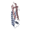
- PDB-4jnu: Crystal structure of the human Nup57CCS3* coiled-coil segment, sp... -
+ Open data

- Basic information
| Entry | Database: PDB / ID: 4jnu | ||||||
|---|---|---|---|---|---|---|---|
| Title | Crystal structure of the human Nup57CCS3* coiled-coil segment, space group P21 | ||||||
 Components Components | Nucleoporin p54 | ||||||
 Keywords Keywords | TRANSPORT PROTEIN / nucleocytoplasmic transport | ||||||
| Function / homology |  Function and homology information Function and homology informationnuclear pore central transport channel / protein localization to nuclear inner membrane / nuclear pore organization / Nuclear Pore Complex (NPC) Disassembly / Regulation of Glucokinase by Glucokinase Regulatory Protein / Defective TPR may confer susceptibility towards thyroid papillary carcinoma (TPC) / Transport of Ribonucleoproteins into the Host Nucleus / Transport of the SLBP independent Mature mRNA / Transport of the SLBP Dependant Mature mRNA / NS1 Mediated Effects on Host Pathways ...nuclear pore central transport channel / protein localization to nuclear inner membrane / nuclear pore organization / Nuclear Pore Complex (NPC) Disassembly / Regulation of Glucokinase by Glucokinase Regulatory Protein / Defective TPR may confer susceptibility towards thyroid papillary carcinoma (TPC) / Transport of Ribonucleoproteins into the Host Nucleus / Transport of the SLBP independent Mature mRNA / Transport of the SLBP Dependant Mature mRNA / NS1 Mediated Effects on Host Pathways / SUMOylation of SUMOylation proteins / structural constituent of nuclear pore / Transport of Mature mRNA Derived from an Intronless Transcript / Rev-mediated nuclear export of HIV RNA / Nuclear import of Rev protein / SUMOylation of RNA binding proteins / NEP/NS2 Interacts with the Cellular Export Machinery / Transport of Mature mRNA derived from an Intron-Containing Transcript / tRNA processing in the nucleus / Postmitotic nuclear pore complex (NPC) reformation / Viral Messenger RNA Synthesis / nucleocytoplasmic transport / NLS-bearing protein import into nucleus / SUMOylation of ubiquitinylation proteins / Vpr-mediated nuclear import of PICs / SUMOylation of DNA replication proteins / Regulation of HSF1-mediated heat shock response / nuclear pore / mRNA transport / SUMOylation of DNA damage response and repair proteins / SUMOylation of chromatin organization proteins / HCMV Late Events / Transcriptional regulation by small RNAs / ISG15 antiviral mechanism / HCMV Early Events / nuclear envelope / snRNP Assembly / nuclear membrane / SARS-CoV-2 activates/modulates innate and adaptive immune responses Similarity search - Function | ||||||
| Biological species |  Homo sapiens (human) Homo sapiens (human) | ||||||
| Method |  X-RAY DIFFRACTION / SYNCHROTRON / AB INITIO PHASING / Resolution: 1.445 Å X-RAY DIFFRACTION / SYNCHROTRON / AB INITIO PHASING / Resolution: 1.445 Å | ||||||
 Authors Authors | Stuwe, T. / Bley, C.J. / Mayo, D.J. / Hoelz, A. | ||||||
 Citation Citation | Journal: Science / Year: 2015 Title: Architecture of the fungal nuclear pore inner ring complex. Authors: Stuwe, T. / Bley, C.J. / Thierbach, K. / Petrovic, S. / Schilbach, S. / Mayo, D.J. / Perriches, T. / Rundlet, E.J. / Jeon, Y.E. / Collins, L.N. / Huber, F.M. / Lin, D.H. / Paduch, M. / ...Authors: Stuwe, T. / Bley, C.J. / Thierbach, K. / Petrovic, S. / Schilbach, S. / Mayo, D.J. / Perriches, T. / Rundlet, E.J. / Jeon, Y.E. / Collins, L.N. / Huber, F.M. / Lin, D.H. / Paduch, M. / Koide, A. / Lu, V. / Fischer, J. / Hurt, E. / Koide, S. / Kossiakoff, A.A. / Hoelz, A. | ||||||
| History |
|
- Structure visualization
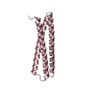
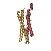
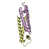
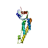
| Structure viewer | Molecule: MolmilJmol/JSmol |
|---|
- Downloads & links
-Download
| PDBx/mmCIF format | 4jnu.cif.gz | 112.8 KB | Display | PDBx/mmCIF format |
|---|---|---|---|---|
| PDB format | pdb4jnu.ent.gz | 93 KB | Display | PDB format |
| PDBx/mmJSON format | 4jnu.json.gz | Tree view | PDBx/mmJSON format | |
| Others |  Other downloads Other downloads |
-Validation report
| Arichive directory | https://data.pdbj.org/pub/pdb/validation_reports/jn/4jnuftp://data.pdbj.org/pub/pdb/validation_reports/jn/4jnu | HTTPS FTP |
|---|
-Related structure data
| Related structure data |  4jnvC  4jo7C  4jo9C  4jq5C  5cwsC 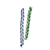 5cwtC  5cwuC  5cwvC  5cwwC C: citing same article ( |
|---|---|
| Similar structure data |


-Links
 PDBj
PDBj


- Assembly
| Deposited unit | 
| ||||||||
|---|---|---|---|---|---|---|---|---|---|
| 1 |
| ||||||||
| Unit cell |
|
-Components
| #1: Protein/peptide | Mass: 4729.410 Da / Num. of mol.: 4 / Fragment: UNP residues 453-491 Source method: isolated from a genetically manipulated source Source: (gene. exp.) Homo sapiens (human) / Gene: NUP54 / Production host:  #2: Water | ChemComp-HOH / |  Mass: 18.015 Da / Num. of mol.: 131 / Source method: isolated from a natural source / Formula: H2O Mass: 18.015 Da / Num. of mol.: 131 / Source method: isolated from a natural source / Formula: H2O |
|---|
-Experimental details
-Experiment
| Experiment | Method: X-RAY DIFFRACTION / Number of used crystals: 1 |
|---|
- Sample preparation
| Crystal | Density Matthews: 2.02 Å3/Da / Density % sol: 39.13 % |
|---|---|
| Crystal grow | Temperature: 294 K / Method: vapor diffusion, hanging drop / pH: 5.3 Details: 0.1 M sodium acetate, pH 5.3, 1.9 M sodium formate, VAPOR DIFFUSION, HANGING DROP, temperature 294K |
-Data collection
| Diffraction | Mean temperature: 100 K |
|---|---|
| Diffraction source | Source: SYNCHROTRON / Site: ALS  / Beamline: 8.2.1 / Wavelength: 1 Å / Beamline: 8.2.1 / Wavelength: 1 Å |
| Detector | Type: ADSC QUANTUM 315r / Detector: CCD / Date: Nov 16, 2012 |
| Radiation | Monochromator: double crystal Si(111) / Protocol: SINGLE WAVELENGTH / Monochromatic (M) / Laue (L): M / Scattering type: x-ray |
| Radiation wavelength | Wavelength: 1 Å / Relative weight: 1 |
| Reflection | Resolution: 1.445→20 Å / Num. obs: 26320 / Observed criterion σ(F): 0 / Biso Wilson estimate: 17.63 Å2 |
- Processing
| Software |
| |||||||||||||||||||||||||||||||||||||||||||||||||||||||||||||||||||||||||||||||||||||||||||||||||||||||||
|---|---|---|---|---|---|---|---|---|---|---|---|---|---|---|---|---|---|---|---|---|---|---|---|---|---|---|---|---|---|---|---|---|---|---|---|---|---|---|---|---|---|---|---|---|---|---|---|---|---|---|---|---|---|---|---|---|---|---|---|---|---|---|---|---|---|---|---|---|---|---|---|---|---|---|---|---|---|---|---|---|---|---|---|---|---|---|---|---|---|---|---|---|---|---|---|---|---|---|---|---|---|---|---|---|---|---|
| Refinement | Method to determine structure: AB INITIO PHASING / Resolution: 1.445→19.946 Å / Occupancy max: 1 / Occupancy min: 0.23 / FOM work R set: 0.8461 / SU ML: 0.14 / σ(F): 1.35 / Phase error: 22.65 / Stereochemistry target values: ML
| |||||||||||||||||||||||||||||||||||||||||||||||||||||||||||||||||||||||||||||||||||||||||||||||||||||||||
| Solvent computation | Shrinkage radii: 0.9 Å / VDW probe radii: 1.11 Å / Solvent model: FLAT BULK SOLVENT MODEL | |||||||||||||||||||||||||||||||||||||||||||||||||||||||||||||||||||||||||||||||||||||||||||||||||||||||||
| Displacement parameters | Biso max: 103.18 Å2 / Biso mean: 27.8702 Å2 / Biso min: 8.65 Å2 | |||||||||||||||||||||||||||||||||||||||||||||||||||||||||||||||||||||||||||||||||||||||||||||||||||||||||
| Refinement step | Cycle: LAST / Resolution: 1.445→19.946 Å
| |||||||||||||||||||||||||||||||||||||||||||||||||||||||||||||||||||||||||||||||||||||||||||||||||||||||||
| Refine LS restraints |
| |||||||||||||||||||||||||||||||||||||||||||||||||||||||||||||||||||||||||||||||||||||||||||||||||||||||||
| LS refinement shell | Refine-ID: X-RAY DIFFRACTION / Total num. of bins used: 14
|
