 Movie
Movie Controller


+ Open data

- Basic information
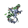
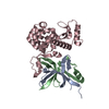
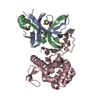
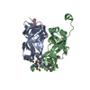
| Entry | Database: PDB / ID: 4yx5 | ||||||
|---|---|---|---|---|---|---|---|
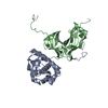
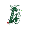
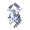
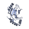
| Title | SpaO(SPOA1,2) | ||||||
 Components Components | (Surface presentation of antigens protein SpaO) x 2 | ||||||
 Keywords Keywords | PROTEIN TRANSPORT / Type III Secretion System | ||||||
| Function / homology |  Function and homology information Function and homology informationprotein secretion by the type III secretion system / bacterial-type flagellum-dependent swarming motility / positive chemotaxis Similarity search - Function | ||||||
| Biological species |  Salmonella typhimurium (bacteria) Salmonella typhimurium (bacteria) | ||||||
| Method |  X-RAY DIFFRACTION / SYNCHROTRON / SAD / Resolution: 2.9001 Å X-RAY DIFFRACTION / SYNCHROTRON / SAD / Resolution: 2.9001 Å | ||||||
 Authors Authors | Notti, R.Q. / Stebbins, C.E. | ||||||
 Citation Citation | Journal: Nat Commun / Year: 2015 Title: A common assembly module in injectisome and flagellar type III secretion sorting platforms. Authors: Notti, R.Q. / Bhattacharya, S. / Lilic, M. / Stebbins, C.E. | ||||||
| History |
|
- Structure visualization
| Structure viewer | Molecule: MolmilJmol/JSmol |
|---|
- Downloads & links
-Download
| PDBx/mmCIF format | 4yx5.cif.gz | 40.1 KB | Display | PDBx/mmCIF format |
|---|---|---|---|---|
| PDB format | pdb4yx5.ent.gz | 26.5 KB | Display | PDB format |
| PDBx/mmJSON format | 4yx5.json.gz | Tree view | PDBx/mmJSON format | |
| Others |  Other downloads Other downloads |
-Validation report
| Summary document | 4yx5_validation.pdf.gz | 428.5 KB | Display | wwPDB validaton report |
|---|---|---|---|---|
| Full document | 4yx5_full_validation.pdf.gz | 430.6 KB | Display | |
| Data in XML | 4yx5_validation.xml.gz | 7.4 KB | Display | |
| Data in CIF | 4yx5_validation.cif.gz | 8.8 KB | Display | |
| Arichive directory | https://data.pdbj.org/pub/pdb/validation_reports/yx/4yx5ftp://data.pdbj.org/pub/pdb/validation_reports/yx/4yx5 | HTTPS FTP |
-Related structure data


-Links
 PDBj
PDBj- Assembly
| Deposited unit | 
| ||||||||
|---|---|---|---|---|---|---|---|---|---|
| 1 |
| ||||||||
| Unit cell |
| ||||||||
| Components on special symmetry positions |
|
-Components
| #1: Protein | Mass: 8246.572 Da / Num. of mol.: 1 / Fragment: UNP Residues 145-213 Source method: isolated from a genetically manipulated source Source: (gene. exp.) Salmonella typhimurium (strain LT2 / SGSC1412 / ATCC 700720) (bacteria)Strain: LT2 / SGSC1412 / ATCC 700720 / Gene: spaO, STM2891 / Production host: |
|---|---|
| #2: Protein | Mass: 7710.809 Da / Num. of mol.: 1 / Fragment: UNP Residues 232-297 Source method: isolated from a genetically manipulated source Source: (gene. exp.) Salmonella typhimurium (bacteria) / Strain: LT2 / SGSC1412 / ATCC 700720 / Gene: spaO, STM2891 / Production host: |
| #3: Chemical | ChemComp-CL /   Mass: 35.453 Da / Num. of mol.: 1 / Source method: obtained synthetically / Formula: Cl Mass: 35.453 Da / Num. of mol.: 1 / Source method: obtained synthetically / Formula: Cl |
-Experimental details
-Experiment
| Experiment | Method: X-RAY DIFFRACTION / Number of used crystals: 1 |
|---|
- Sample preparation
| Crystal | Density Matthews: 3.44 Å3/Da / Density % sol: 64.26 % |
|---|---|
| Crystal grow | Temperature: 277 K / Method: vapor diffusion, hanging drop Details: SpaO(145-213) + SpaO (232-297) was concentrated to 12mg/mL and crystallized with 25% PEG400, 10% isopropanol, 100mM sodium citrate pH=5.6 at 277K. Microseeding was employed to enhance ...Details: SpaO(145-213) + SpaO (232-297) was concentrated to 12mg/mL and crystallized with 25% PEG400, 10% isopropanol, 100mM sodium citrate pH=5.6 at 277K. Microseeding was employed to enhance crystal uniformity and diffraction. Briefly, crystals to be seeded were harvested in precipitant solution and vortexed in a microfuge tube with a small stir bar for ~60 seconds. The slurry of microseeds was serially dilluted (5-10-fold steps) in precipitant solution and 5 selected microseed-precipitant mixtures were mixed with fresh protein as in a normal hanging drop experiment. Crystals were cryoprotected in mother liquor with the PEG400 concentration raised to 37.5%. |
-Data collection
| Diffraction | Mean temperature: 100 K | |||||||||||||||||||||||||||
|---|---|---|---|---|---|---|---|---|---|---|---|---|---|---|---|---|---|---|---|---|---|---|---|---|---|---|---|---|
| Diffraction source | Source: SYNCHROTRON / Site: NSLS  / Beamline: X29A / Wavelength: 1.075, 0.979 / Beamline: X29A / Wavelength: 1.075, 0.979 | |||||||||||||||||||||||||||
| Detector | Type: ADSC QUANTUM 315r / Detector: CCD / Date: Nov 22, 2013 | |||||||||||||||||||||||||||
| Radiation | Protocol: SINGLE WAVELENGTH / Monochromatic (M) / Laue (L): M / Scattering type: x-ray | |||||||||||||||||||||||||||
| Radiation wavelength |
| |||||||||||||||||||||||||||
| Reflection | Resolution: 2.9→54.19 Å / Num. obs: 4975 / % possible obs: 99.3 % / Redundancy: 24.6 % / CC1/2: 0.996 / Rmerge(I) obs: 0.166 / Rpim(I) all: 0.034 / Net I/σ(I): 14.4 / Num. measured all: 122494 / Scaling rejects: 9 | |||||||||||||||||||||||||||
| Reflection shell | Diffraction-ID: 1 / Rejects: _
|
- Processing
| Software |
| ||||||||||||||||||||||||||||||
|---|---|---|---|---|---|---|---|---|---|---|---|---|---|---|---|---|---|---|---|---|---|---|---|---|---|---|---|---|---|---|---|
| Refinement | Method to determine structure: SAD / Resolution: 2.9001→38.678 Å / SU ML: 0.46 / Cross valid method: FREE R-VALUE / σ(F): 1.39 / Phase error: 28.3 / Stereochemistry target values: ML
| ||||||||||||||||||||||||||||||
| Solvent computation | Shrinkage radii: 0.9 Å / VDW probe radii: 1.11 Å / Solvent model: FLAT BULK SOLVENT MODEL | ||||||||||||||||||||||||||||||
| Displacement parameters | Biso max: 154.08 Å2 / Biso mean: 74.2199 Å2 / Biso min: 37.22 Å2 | ||||||||||||||||||||||||||||||
| Refinement step | Cycle: final / Resolution: 2.9001→38.678 Å
| ||||||||||||||||||||||||||||||
| Refine LS restraints |
| ||||||||||||||||||||||||||||||
| LS refinement shell | Refine-ID: X-RAY DIFFRACTION / Total num. of bins used: 4 / % reflection obs: 99 %
|
