 Movie
Movie Controller

[English] 日本語
 Yorodumi
- PDB-1i7k: CRYSTAL STRUCTURE OF HUMAN MITOTIC-SPECIFIC UBIQUITIN-CONJUGATING... -
+ Open data

- Basic information
| Entry | Database: PDB / ID: 1i7k | ||||||
|---|---|---|---|---|---|---|---|
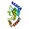
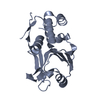
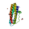
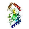
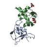
| Title | CRYSTAL STRUCTURE OF HUMAN MITOTIC-SPECIFIC UBIQUITIN-CONJUGATING ENZYME, UBCH10 | ||||||
 Components Components | UBIQUITIN-CONJUGATING ENZYME E2 H10 | ||||||
 Keywords Keywords | LIGASE / ubiquitin conjugating enzyme | ||||||
| Function / homology |  Function and homology information Function and homology informationpositive regulation of ubiquitin protein ligase activity / free ubiquitin chain polymerization / positive regulation of exit from mitosis / Conversion from APC/C:Cdc20 to APC/C:Cdh1 in late anaphase / Inactivation of APC/C via direct inhibition of the APC/C complex / APC/C:Cdc20 mediated degradation of mitotic proteins / anaphase-promoting complex / anaphase-promoting complex-dependent catabolic process / Aberrant regulation of mitotic exit in cancer due to RB1 defects / Phosphorylation of the APC/C ...positive regulation of ubiquitin protein ligase activity / free ubiquitin chain polymerization / positive regulation of exit from mitosis / Conversion from APC/C:Cdc20 to APC/C:Cdh1 in late anaphase / Inactivation of APC/C via direct inhibition of the APC/C complex / APC/C:Cdc20 mediated degradation of mitotic proteins / anaphase-promoting complex / anaphase-promoting complex-dependent catabolic process / Aberrant regulation of mitotic exit in cancer due to RB1 defects / Phosphorylation of the APC/C / (E3-independent) E2 ubiquitin-conjugating enzyme / positive regulation of mitotic metaphase/anaphase transition / exit from mitosis / protein K11-linked ubiquitination / regulation of mitotic metaphase/anaphase transition / E2 ubiquitin-conjugating enzyme / Regulation of APC/C activators between G1/S and early anaphase / ubiquitin conjugating enzyme activity / Transcriptional Regulation by VENTX / ubiquitin-like protein ligase binding / ubiquitin ligase complex / protein K48-linked ubiquitination / APC/C:Cdc20 mediated degradation of Cyclin B / APC-Cdc20 mediated degradation of Nek2A / Synthesis of active ubiquitin: roles of E1 and E2 enzymes / Autodegradation of Cdh1 by Cdh1:APC/C / APC/C:Cdc20 mediated degradation of Securin / Assembly of the pre-replicative complex / Cdc20:Phospho-APC/C mediated degradation of Cyclin A / APC/C:Cdh1 mediated degradation of Cdc20 and other APC/C:Cdh1 targeted proteins in late mitosis/early G1 / CDK-mediated phosphorylation and removal of Cdc6 / protein polyubiquitination / ubiquitin-protein transferase activity / Separation of Sister Chromatids / ubiquitin protein ligase activity / Antigen processing: Ubiquitination & Proteasome degradation / Senescence-Associated Secretory Phenotype (SASP) / ubiquitin-dependent protein catabolic process / proteasome-mediated ubiquitin-dependent protein catabolic process / protein ubiquitination / cell division / nucleoplasm / ATP binding / nucleus / plasma membrane / cytosol Similarity search - Function | ||||||
| Biological species |  Homo sapiens (human) Homo sapiens (human) | ||||||
| Method |  X-RAY DIFFRACTION / MOLECULAR REPLACEMENT / Resolution: 1.95 Å X-RAY DIFFRACTION / MOLECULAR REPLACEMENT / Resolution: 1.95 Å | ||||||
 Authors Authors | Basavappa, R. / Lin, Y. | ||||||
 Citation Citation | Journal: J.Biol.Chem. / Year: 2002 Title: Structural and functional analysis of the human mitotic-specific ubiquitin-conjugating enzyme, UbcH10. Authors: Lin, Y. / Hwang, W.C. / Basavappa, R. | ||||||
| History |
|
- Structure visualization
| Structure viewer | Molecule: MolmilJmol/JSmol |
|---|
- Downloads & links
-Download
| PDBx/mmCIF format | 1i7k.cif.gz | 74.2 KB | Display | PDBx/mmCIF format |
|---|---|---|---|---|
| PDB format | pdb1i7k.ent.gz | 55.5 KB | Display | PDB format |
| PDBx/mmJSON format | 1i7k.json.gz | Tree view | PDBx/mmJSON format | |
| Others |  Other downloads Other downloads |
-Validation report
| Arichive directory | https://data.pdbj.org/pub/pdb/validation_reports/i7/1i7kftp://data.pdbj.org/pub/pdb/validation_reports/i7/1i7k | HTTPS FTP |
|---|
-Related structure data
| Related structure data |  2e2cS S: Starting model for refinement |
|---|---|
| Similar structure data |


-Links
 PDBj
PDBj


- Assembly
| Deposited unit | 
| ||||||||
|---|---|---|---|---|---|---|---|---|---|
| 1 | 
| ||||||||
| 2 | 
| ||||||||
| Unit cell |
|
-Components
| #1: Protein | Mass: 19657.197 Da / Num. of mol.: 2 / Mutation: C114S Source method: isolated from a genetically manipulated source Source: (gene. exp.) Homo sapiens (human) / Gene: UBCH10 / Plasmid: PT7-7 / Production host:  #2: Water | ChemComp-HOH / |  Mass: 18.015 Da / Num. of mol.: 196 / Source method: isolated from a natural source / Formula: H2O Mass: 18.015 Da / Num. of mol.: 196 / Source method: isolated from a natural source / Formula: H2O |
|---|
-Experimental details
-Experiment
| Experiment | Method: X-RAY DIFFRACTION / Number of used crystals: 2 |
|---|
- Sample preparation
| Crystal | Density Matthews: 2.24 Å3/Da / Density % sol: 45.06 % | |||||||||||||||
|---|---|---|---|---|---|---|---|---|---|---|---|---|---|---|---|---|
| Crystal grow | Temperature: 293 K / Method: vapor diffusion, hanging drop / pH: 7 Details: PEG 1500, sodium chloride, sodium acetate, pH 7.0, VAPOR DIFFUSION, HANGING DROP, temperature 293K | |||||||||||||||
| Crystal grow | *PLUS Temperature: 20 ℃ / Method: vapor diffusionDetails: drop consists of equal amounts of protein and precipitant solutions | |||||||||||||||
| Components of the solutions | *PLUS
|
-Data collection
| Diffraction | Mean temperature: 293 K |
|---|---|
| Diffraction source | Source: ROTATING ANODE / Type: RIGAKU RU200 / Wavelength: 1.5418 Å |
| Detector | Type: RIGAKU RAXIS IV / Detector: IMAGE PLATE / Date: Jun 29, 2000 / Details: Osmic mirrors |
| Radiation | Monochromator: Osmic mirrors / Protocol: SINGLE WAVELENGTH / Monochromatic (M) / Laue (L): M / Scattering type: x-ray |
| Radiation wavelength | Wavelength: 1.5418 Å / Relative weight: 1 |
| Reflection | Resolution: 1.94→50 Å / Num. all: 24518 / Num. obs: 24518 / % possible obs: 97.7 % / Observed criterion σ(I): -3 / Redundancy: 2.9 % / Rmerge(I) obs: 0.061 / Net I/σ(I): 25.6 |
| Reflection shell | Resolution: 1.94→2.01 Å / Redundancy: 2.532 % / Rmerge(I) obs: 0.319 / Mean I/σ(I) obs: 4.167 / Num. unique all: 2218 / % possible all: 88.8 |
| Reflection shell | *PLUS % possible obs: 88.8 % / Num. unique obs: 2218 |
- Processing
| Software |
| |||||||||||||||||||||||||
|---|---|---|---|---|---|---|---|---|---|---|---|---|---|---|---|---|---|---|---|---|---|---|---|---|---|---|
| Refinement | Method to determine structure: MOLECULAR REPLACEMENT Starting model: PDB ENTRY 2E2C Resolution: 1.95→50 Å / Isotropic thermal model: isotropic / Cross valid method: THROUGHOUT / σ(F): 0 / σ(I): 0 / Stereochemistry target values: Engh & Huber
| |||||||||||||||||||||||||
| Displacement parameters | Biso mean: 35.808 Å2 | |||||||||||||||||||||||||
| Refinement step | Cycle: LAST / Resolution: 1.95→50 Å
| |||||||||||||||||||||||||
| Refine LS restraints |
| |||||||||||||||||||||||||
| Software | *PLUS Name: CNS / Classification: refinement | |||||||||||||||||||||||||
| Refinement | *PLUS Rfactor Rfree: 0.226 / Rfactor Rwork: 0.176 | |||||||||||||||||||||||||
| Solvent computation | *PLUS | |||||||||||||||||||||||||
| Displacement parameters | *PLUS | |||||||||||||||||||||||||
| Refine LS restraints | *PLUS
|
