 Movie
Movie Controller

[English] 日本語
 Yorodumi
- PDB-2e2c: E2-C, AN UBIQUITIN CONJUGATING ENZYME REQUIRED FOR THE DESTRUCTIO... -
+ Open data

- Basic information
| Entry | Database: PDB / ID: 2e2c | |||||||||
|---|---|---|---|---|---|---|---|---|---|---|
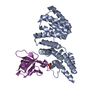
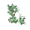
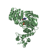
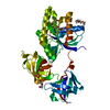
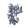
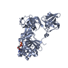
| Title | E2-C, AN UBIQUITIN CONJUGATING ENZYME REQUIRED FOR THE DESTRUCTION OF MITOTIC CYCLINS | |||||||||
 Components Components | UBIQUITIN CONJUGATING ENZYME | |||||||||
 Keywords Keywords | UBIQUITIN CONJUGATION / UBIQUITIN CARRIER PROTEIN / THIOESTER BOND / LIGASE | |||||||||
| Function / homology |  Function and homology information Function and homology information(E3-independent) E2 ubiquitin-conjugating enzyme / E2 ubiquitin-conjugating enzyme / ubiquitin conjugating enzyme activity / protein ubiquitination / cell division / ATP binding Similarity search - Function | |||||||||
| Biological species |  Spisula solidissima (Atlantic surf clam) Spisula solidissima (Atlantic surf clam) | |||||||||
| Method |  X-RAY DIFFRACTION / MOLECULAR REPLACEMENT / Resolution: 2 Å X-RAY DIFFRACTION / MOLECULAR REPLACEMENT / Resolution: 2 Å | |||||||||
 Authors Authors | Jiang, F. / Basavappa, R. | |||||||||
 Citation Citation | Journal: Biochemistry / Year: 1999 Title: Crystal structure of the cyclin-specific ubiquitin-conjugating enzyme from clam, E2-C, at 2.0 A resolution. Authors: Jiang, F. / Basavappa, R. | |||||||||
| History |
|
- Structure visualization
| Structure viewer | Molecule: MolmilJmol/JSmol |
|---|
- Downloads & links
-Download
| PDBx/mmCIF format | 2e2c.cif.gz | 47.1 KB | Display | PDBx/mmCIF format |
|---|---|---|---|---|
| PDB format | pdb2e2c.ent.gz | 32.9 KB | Display | PDB format |
| PDBx/mmJSON format | 2e2c.json.gz | Tree view | PDBx/mmJSON format | |
| Others |  Other downloads Other downloads |
-Validation report
| Arichive directory | https://data.pdbj.org/pub/pdb/validation_reports/e2/2e2cftp://data.pdbj.org/pub/pdb/validation_reports/e2/2e2c | HTTPS FTP |
|---|
-Related structure data
| Related structure data |  1aak S: Starting model for refinement |
|---|---|
| Similar structure data |


-Links
 PDBj
PDBj

- Assembly
| Deposited unit | 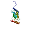
| ||||||||
|---|---|---|---|---|---|---|---|---|---|
| 1 | 
| ||||||||
| Unit cell |
| ||||||||
| Components on special symmetry positions |
|
-Components
| #1: Protein | Mass: 17762.057 Da / Num. of mol.: 1 / Source method: isolated from a natural source / Source: (natural) Spisula solidissima (Atlantic surf clam) / References: UniProt: Q95044, ubiquitin-protein ligase |
|---|---|
| #2: Water | ChemComp-HOH /  Mass: 18.015 Da / Num. of mol.: 172 / Source method: isolated from a natural source / Formula: H2O Mass: 18.015 Da / Num. of mol.: 172 / Source method: isolated from a natural source / Formula: H2O |
-Experimental details
-Experiment
| Experiment | Method: X-RAY DIFFRACTION / Number of used crystals: 1 |
|---|
- Sample preparation
| Crystal | Density Matthews: 2.64 Å3/Da / Density % sol: 53 % | ||||||||||||||||||||
|---|---|---|---|---|---|---|---|---|---|---|---|---|---|---|---|---|---|---|---|---|---|
| Crystal grow | pH: 7.4 / Details: pH 7.4 | ||||||||||||||||||||
| Crystal grow | *PLUS pH: 8.5 / Method: vapor diffusion, hanging drop | ||||||||||||||||||||
| Components of the solutions | *PLUS
|
-Data collection
| Diffraction | Mean temperature: 103 K |
|---|---|
| Diffraction source | Source: ROTATING ANODE / Type: RIGAKU RUH2R / Wavelength: 1.5418 |
| Detector | Type: RIGAKU / Detector: IMAGE PLATE / Date: Oct 1, 1996 / Details: DOUBLE MIRRORS |
| Radiation | Monochromator: NI FILTER / Monochromatic (M) / Laue (L): M / Scattering type: x-ray |
| Radiation wavelength | Wavelength: 1.5418 Å / Relative weight: 1 |
| Reflection | Resolution: 2→50 Å / Num. obs: 13327 / % possible obs: 99.4 % / Observed criterion σ(I): 2 / Redundancy: 4 % / Biso Wilson estimate: 14 Å2 / Rmerge(I) obs: 0.061 / Net I/σ(I): 11.7 |
| Reflection shell | Resolution: 2→2.2 Å / Redundancy: 2 % / Rmerge(I) obs: 0.36 / Mean I/σ(I) obs: 3.7 / % possible all: 92.7 |
| Reflection shell | *PLUS % possible obs: 92.7 % / Rmerge(I) obs: 0.358 |
- Processing
| Software |
| ||||||||||||||||||||||||||||||||||||||||||||||||||||||||||||
|---|---|---|---|---|---|---|---|---|---|---|---|---|---|---|---|---|---|---|---|---|---|---|---|---|---|---|---|---|---|---|---|---|---|---|---|---|---|---|---|---|---|---|---|---|---|---|---|---|---|---|---|---|---|---|---|---|---|---|---|---|---|
| Refinement | Method to determine structure: MOLECULAR REPLACEMENT Starting model: 1AAK 1aak Resolution: 2→20 Å / Cross valid method: THROUGHOUT / σ(F): 0
| ||||||||||||||||||||||||||||||||||||||||||||||||||||||||||||
| Displacement parameters | Biso mean: 27 Å2 | ||||||||||||||||||||||||||||||||||||||||||||||||||||||||||||
| Refinement step | Cycle: LAST / Resolution: 2→20 Å
| ||||||||||||||||||||||||||||||||||||||||||||||||||||||||||||
| Refine LS restraints |
| ||||||||||||||||||||||||||||||||||||||||||||||||||||||||||||
| Software | *PLUS Name: X-PLOR / Version: 3.851 / Classification: refinement | ||||||||||||||||||||||||||||||||||||||||||||||||||||||||||||
| Refinement | *PLUS Rfactor Rfree: 0.297 | ||||||||||||||||||||||||||||||||||||||||||||||||||||||||||||
| Solvent computation | *PLUS | ||||||||||||||||||||||||||||||||||||||||||||||||||||||||||||
| Displacement parameters | *PLUS | ||||||||||||||||||||||||||||||||||||||||||||||||||||||||||||
| Refine LS restraints | *PLUS
|
