 Movie
Movie Controller


+ Open data

- Basic information
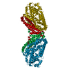
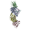
| Entry | Database: PDB / ID: 4y5y | ||||||
|---|---|---|---|---|---|---|---|
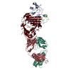
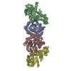
| Title | Diabody 330 complex with EpoR | ||||||
 Components Components |
| ||||||
 Keywords Keywords | protein binding/immune system / diabody complex / receptor / protein binding-immune system complex | ||||||
| Function / homology |  Function and homology information Function and homology informationerythropoietin receptor activity / Signaling by Erythropoietin / Erythropoietin activates STAT5 / Erythropoietin activates Phospholipase C gamma (PLCG) / erythropoietin-mediated signaling pathway / Erythropoietin activates Phosphoinositide-3-kinase (PI3K) / decidualization / Erythropoietin activates RAS / brain development / heart development ...erythropoietin receptor activity / Signaling by Erythropoietin / Erythropoietin activates STAT5 / Erythropoietin activates Phospholipase C gamma (PLCG) / erythropoietin-mediated signaling pathway / Erythropoietin activates Phosphoinositide-3-kinase (PI3K) / decidualization / Erythropoietin activates RAS / brain development / heart development / nuclear speck / external side of plasma membrane / signal transduction / extracellular region / identical protein binding / plasma membrane Similarity search - Function | ||||||
| Biological species |  Homo sapiens (human) Homo sapiens (human) | ||||||
| Method |  X-RAY DIFFRACTION / SYNCHROTRON / MOLECULAR REPLACEMENT / Resolution: 2.85 Å X-RAY DIFFRACTION / SYNCHROTRON / MOLECULAR REPLACEMENT / Resolution: 2.85 Å | ||||||
 Authors Authors | Moraga, I. / Guo, F. / Ozkan, E. / Jude, K.M. / Garcia, K.C. | ||||||
 Citation Citation | Journal: Cell / Year: 2015 Title: Tuning Cytokine Receptor Signaling by Re-orienting Dimer Geometry with Surrogate Ligands. Authors: Moraga, I. / Wernig, G. / Wilmes, S. / Gryshkova, V. / Richter, C.P. / Hong, W.J. / Sinha, R. / Guo, F. / Fabionar, H. / Wehrman, T.S. / Krutzik, P. / Demharter, S. / Plo, I. / Weissman, I.L. ...Authors: Moraga, I. / Wernig, G. / Wilmes, S. / Gryshkova, V. / Richter, C.P. / Hong, W.J. / Sinha, R. / Guo, F. / Fabionar, H. / Wehrman, T.S. / Krutzik, P. / Demharter, S. / Plo, I. / Weissman, I.L. / Minary, P. / Majeti, R. / Constantinescu, S.N. / Piehler, J. / Garcia, K.C. | ||||||
| History |
|
- Structure visualization
| Structure viewer | Molecule: MolmilJmol/JSmol |
|---|
- Downloads & links
-Download
| PDBx/mmCIF format | 4y5y.cif.gz | 336.3 KB | Display | PDBx/mmCIF format |
|---|---|---|---|---|
| PDB format | pdb4y5y.ent.gz | 274.2 KB | Display | PDB format |
| PDBx/mmJSON format | 4y5y.json.gz | Tree view | PDBx/mmJSON format | |
| Others |  Other downloads Other downloads |
-Validation report
| Arichive directory | https://data.pdbj.org/pub/pdb/validation_reports/y5/4y5yftp://data.pdbj.org/pub/pdb/validation_reports/y5/4y5y | HTTPS FTP |
|---|
-Related structure data
| Related structure data |  4y5vC  4y5xC 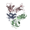 1eerS 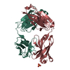 3s34S C: citing same article ( S: Starting model for refinement |
|---|---|
| Similar structure data |


-Links
 PDBj
PDBj


- Assembly
| Deposited unit | 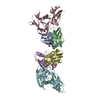
| ||||||||
|---|---|---|---|---|---|---|---|---|---|
| 1 |
| ||||||||
| Unit cell |
| ||||||||
| Details | biological unit is the same as asymmetric unit as confirmed by beta-gal complementation, signalling assay, superresolution microscopy, MALS |
-Components
| #1: Antibody | Mass: 13941.515 Da / Num. of mol.: 2 Source method: isolated from a genetically manipulated source Source: (gene. exp.) Homo sapiens (human) / Production host:  Trichoplusia ni (cabbage looper) Trichoplusia ni (cabbage looper)#2: Antibody | Mass: 12016.095 Da / Num. of mol.: 2 Source method: isolated from a genetically manipulated source Source: (gene. exp.) Homo sapiens (human) / Production host: Trichoplusia ni (cabbage looper)#3: Protein | Mass: 25210.562 Da / Num. of mol.: 2 / Mutation: N76Q, N188Q, D247L, and L248D Source method: isolated from a genetically manipulated source Source: (gene. exp.) Homo sapiens (human) / Gene: EPOR / Production host: Trichoplusia ni (cabbage looper) / References: UniProt: P19235#4: Chemical | ChemComp-GOL / |   Mass: 92.094 Da / Num. of mol.: 1 / Source method: obtained synthetically / Formula: C3H8O3 Mass: 92.094 Da / Num. of mol.: 1 / Source method: obtained synthetically / Formula: C3H8O3#5: Water | ChemComp-HOH / |  Mass: 18.015 Da / Num. of mol.: 33 / Source method: isolated from a natural source / Formula: H2O Mass: 18.015 Da / Num. of mol.: 33 / Source method: isolated from a natural source / Formula: H2O |
|---|
-Experimental details
-Experiment
| Experiment | Method: X-RAY DIFFRACTION / Number of used crystals: 1 |
|---|
- Sample preparation
| Crystal | Density Matthews: 2.66 Å3/Da / Density % sol: 53.73 % |
|---|---|
| Crystal grow | Temperature: 293 K / Method: vapor diffusion / pH: 7 / Details: Magnesium chloride, Tris, PEG 8000 |
-Data collection
| Diffraction | Mean temperature: 100 K | |||||||||||||||||||||||||||
|---|---|---|---|---|---|---|---|---|---|---|---|---|---|---|---|---|---|---|---|---|---|---|---|---|---|---|---|---|
| Diffraction source | Source: SYNCHROTRON / Site: SSRL  / Beamline: BL12-2 / Wavelength: 0.9795 Å / Beamline: BL12-2 / Wavelength: 0.9795 Å | |||||||||||||||||||||||||||
| Detector | Type: PSI PILATUS 6M / Detector: PIXEL / Date: Jun 6, 2013 | |||||||||||||||||||||||||||
| Radiation | Protocol: SINGLE WAVELENGTH / Monochromatic (M) / Laue (L): M / Scattering type: x-ray | |||||||||||||||||||||||||||
| Radiation wavelength | Wavelength: 0.9795 Å / Relative weight: 1 | |||||||||||||||||||||||||||
| Reflection | Resolution: 2.85→48.32 Å / Num. obs: 25898 / % possible obs: 99.4 % / Redundancy: 4.3 % / Biso Wilson estimate: 62.43 Å2 / CC1/2: 0.997 / Rmerge(I) obs: 0.1 / Rpim(I) all: 0.054 / Net I/σ(I): 11.1 / Num. measured all: 110916 | |||||||||||||||||||||||||||
| Reflection shell | Diffraction-ID: 1 / Rejects: _
|
- Processing
| Software |
| ||||||||||||||||||||||||||||||||||||||||||
|---|---|---|---|---|---|---|---|---|---|---|---|---|---|---|---|---|---|---|---|---|---|---|---|---|---|---|---|---|---|---|---|---|---|---|---|---|---|---|---|---|---|---|---|
| Refinement | Method to determine structure: MOLECULAR REPLACEMENT Starting model: 3S34, 1EER Resolution: 2.85→43.146 Å / Cross valid method: FREE R-VALUE
| ||||||||||||||||||||||||||||||||||||||||||
| Solvent computation | Shrinkage radii: 0.9 Å / VDW probe radii: 1.11 Å | ||||||||||||||||||||||||||||||||||||||||||
| Refine analyze | Luzzati sigma a obs: 0.36 Å | ||||||||||||||||||||||||||||||||||||||||||
| Refinement step | Cycle: LAST / Resolution: 2.85→43.146 Å
| ||||||||||||||||||||||||||||||||||||||||||
| LS refinement shell | Resolution: 2.85→2.9048 Å /
| ||||||||||||||||||||||||||||||||||||||||||
| Refinement TLS params. | Method: refined / Origin x: 11.7429 Å / Origin y: 22.1547 Å / Origin z: -53.5305 Å
| ||||||||||||||||||||||||||||||||||||||||||
| Refinement TLS group |
|
