 Movie
Movie Controller

[English] 日本語
 Yorodumi
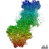
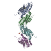
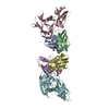
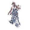
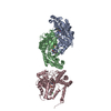
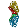
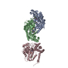
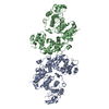
- PDB-3bt2: Structure of urokinase receptor, urokinase and vitronectin complex -
+ Open data

- Basic information
| Entry | Database: PDB / ID: 3bt2 | |||||||||
|---|---|---|---|---|---|---|---|---|---|---|
| Title | Structure of urokinase receptor, urokinase and vitronectin complex | |||||||||
 Components Components |
| |||||||||
 Keywords Keywords | IMMUNE SYSTEM / protein-protein interaction / Glycoprotein / GPI-anchor / Lipoprotein / Membrane / Receptor / Secreted / Blood coagulation / EGF-like domain / Fibrinolysis / Hydrolase / Kringle / Phosphoprotein / Plasminogen activation / Protease / Serine protease / Zymogen / Cell adhesion / Heparin-binding / Sulfation / Immunoglobulin domain | |||||||||
| Function / homology |  Function and homology information Function and homology informationurokinase plasminogen activator receptor activity / Attachment of GPI anchor to uPAR / symbiont-containing vacuole / smooth muscle cell-matrix adhesion / rough endoplasmic reticulum lumen / peptidase inhibitor complex / positive regulation of integrin-mediated signaling pathway / u-plasminogen activator / regulation of smooth muscle cell-matrix adhesion / urokinase plasminogen activator signaling pathway ...urokinase plasminogen activator receptor activity / Attachment of GPI anchor to uPAR / symbiont-containing vacuole / smooth muscle cell-matrix adhesion / rough endoplasmic reticulum lumen / peptidase inhibitor complex / positive regulation of integrin-mediated signaling pathway / u-plasminogen activator / regulation of smooth muscle cell-matrix adhesion / urokinase plasminogen activator signaling pathway / regulation of plasminogen activation / positive regulation of homotypic cell-cell adhesion / regulation of integrin-mediated signaling pathway / alphav-beta3 integrin-vitronectin complex / regulation of fibrinolysis / protein complex involved in cell-matrix adhesion / regulation of wound healing / negative regulation of plasminogen activation / positive regulation of cell-substrate adhesion / positive regulation of vascular endothelial growth factor signaling pathway / serine-type endopeptidase complex / regulation of smooth muscle cell migration / Dissolution of Fibrin Clot / regulation of cell adhesion mediated by integrin / extracellular matrix binding / positive regulation of vascular endothelial growth factor receptor signaling pathway / scavenger receptor activity / extrinsic component of membrane / cell adhesion mediated by integrin / positive regulation of DNA binding / Molecules associated with elastic fibres / smooth muscle cell migration / plasminogen activation / positive regulation of smooth muscle cell migration / extracellular matrix structural constituent / Syndecan interactions / positive regulation of wound healing / polysaccharide binding / negative regulation of intrinsic apoptotic signaling pathway / endodermal cell differentiation / positive regulation of release of cytochrome c from mitochondria / oligodendrocyte differentiation / tertiary granule membrane / basement membrane / regulation of proteolysis / negative regulation of blood coagulation / negative regulation of fibrinolysis / protein polymerization / ECM proteoglycans / Integrin cell surface interactions / regulation of cell adhesion / serine protease inhibitor complex / specific granule membrane / fibrinolysis / positive regulation of epidermal growth factor receptor signaling pathway / collagen binding / negative regulation of proteolysis / cell projection / extracellular matrix organization / liver regeneration / Regulation of Complement cascade / cell-matrix adhesion / Developmental Lineage of Pancreatic Ductal Cells / integrin-mediated signaling pathway / positive regulation of receptor-mediated endocytosis / positive regulation of protein phosphorylation / Golgi lumen / integrin binding / chemotaxis / blood coagulation / regulation of cell population proliferation / cell migration / heparin binding / extracellular matrix / signaling receptor activity / blood microparticle / response to hypoxia / cell adhesion / immune response / positive regulation of cell migration / endoplasmic reticulum lumen / receptor ligand activity / signaling receptor binding / serine-type endopeptidase activity / external side of plasma membrane / protein domain specific binding / focal adhesion / Neutrophil degranulation / endoplasmic reticulum membrane / negative regulation of apoptotic process / enzyme binding / cell surface / signal transduction / endoplasmic reticulum / proteolysis / : / extracellular exosome / extracellular region / membrane / identical protein binding Similarity search - Function | |||||||||
| Biological species |  Homo sapiens (human) Homo sapiens (human) | |||||||||
| Method |  X-RAY DIFFRACTION / SYNCHROTRON / MOLECULAR REPLACEMENT / Resolution: 2.5 Å X-RAY DIFFRACTION / SYNCHROTRON / MOLECULAR REPLACEMENT / Resolution: 2.5 Å | |||||||||
 Authors Authors | Huang, M. | |||||||||
 Citation Citation | Journal: Nat.Struct.Mol.Biol. / Year: 2008 Title: Crystal structures of two human vitronectin, urokinase and urokinase receptor complexes Authors: Huai, Q. / Zhou, A. / Lin, L. / Mazar, A.P. / Parry, G.C. / Callahan, J. / Shaw, D.E. / Furie, B. / Furie, B.C. / Huang, M. | |||||||||
| History |
|
- Structure visualization
| Structure viewer | Molecule: MolmilJmol/JSmol |
|---|
- Downloads & links
-Download
| PDBx/mmCIF format | 3bt2.cif.gz | 183 KB | Display | PDBx/mmCIF format |
|---|---|---|---|---|
| PDB format | pdb3bt2.ent.gz | 141.5 KB | Display | PDB format |
| PDBx/mmJSON format | 3bt2.json.gz | Tree view | PDBx/mmJSON format | |
| Others |  Other downloads Other downloads |
-Validation report
| Arichive directory | https://data.pdbj.org/pub/pdb/validation_reports/bt/3bt2ftp://data.pdbj.org/pub/pdb/validation_reports/bt/3bt2 | HTTPS FTP |
|---|
-Related structure data
| Related structure data |  3bt1C 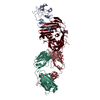 2fd6S S: Starting model for refinement C: citing same article ( |
|---|---|
| Similar structure data |

-Links
 PDBj
PDBj


- Assembly
| Deposited unit | 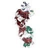
| ||||||||
|---|---|---|---|---|---|---|---|---|---|
| 1 |
| ||||||||
| 2 | 
| ||||||||
| 3 | 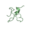
| ||||||||
| Unit cell |
|
-Components
-Protein , 2 types, 2 molecules AU
| #1: Protein | Mass: 15359.318 Da / Num. of mol.: 1 Fragment: urokinase amino terminal fragment, Urokinase-type plasminogen activator long chain A, UNP residues 21-153 Source method: isolated from a genetically manipulated source Source: (gene. exp.) Homo sapiens (human) / Gene: PLAU / Plasmid: pMT/Bip / Species (production host): Escherichia coli / Production host:  |
|---|---|
| #5: Protein | Mass: 31601.479 Da / Num. of mol.: 1 Source method: isolated from a genetically manipulated source Source: (gene. exp.) Homo sapiens (human) / Gene: PLAUR, MO3, UPAR / Plasmid: pMT/Bip / Production host:  |
-Antibody , 2 types, 2 molecules LH
| #3: Antibody | Mass: 23269.691 Da / Num. of mol.: 1 / Fragment: Fab fragment, light chain Source method: isolated from a genetically manipulated source Source: (gene. exp.) |
|---|---|
| #4: Antibody | Mass: 23041.734 Da / Num. of mol.: 1 / Fragment: Fab fragment, heavy chain Source method: isolated from a genetically manipulated source Source: (gene. exp.) |
-Protein/peptide / Non-polymers , 2 types, 58 molecules B
| #2: Protein/peptide | Mass: 4573.103 Da / Num. of mol.: 1 / Fragment: sometomedin-B domain Source method: isolated from a genetically manipulated source Source: (gene. exp.) Homo sapiens (human) / Gene: VTN / Species (production host): Escherichia coli / Production host: |
|---|---|
| #8: Water | ChemComp-HOH / Mass: 18.015 Da / Num. of mol.: 57 / Source method: isolated from a natural source / Formula: H2O |
-Sugars , 2 types, 3 molecules 
| #6: Polysaccharide | Source method: isolated from a genetically manipulated source #7: Sugar | ChemComp-NAG / |  Type: D-saccharide, beta linking / Mass: 221.208 Da / Num. of mol.: 1 Type: D-saccharide, beta linking / Mass: 221.208 Da / Num. of mol.: 1Source method: isolated from a genetically manipulated source Formula: C8H15NO6 |
|---|
-Details
| Has protein modification | Y |
|---|---|
| Sequence details | THIS COORDINATES IN CHAINS L AND H ARE USED NON-SEQUENTIAL RESIDUE NUMBERING. IT IS DUE TO KABAT-WU ...THIS COORDINATE |
-Experimental details
-Experiment
| Experiment | Method: X-RAY DIFFRACTION / Number of used crystals: 2 |
|---|
- Sample preparation
| Crystal | Density Matthews: 2.88 Å3/Da / Density % sol: 57.25 % |
|---|---|
| Crystal grow | Temperature: 295 K / Method: microdialysis / pH: 7.5 Details: 8% PEG 4000, 2.5% ethanol, 0.05% sodium azide, 50mM cacodylate pH 6.5, pH 7.5, MICRODIALYSIS, temperature 295K |
-Data collection
| Diffraction |
| |||||||||||||||
|---|---|---|---|---|---|---|---|---|---|---|---|---|---|---|---|---|
| Diffraction source |
| |||||||||||||||
| Detector |
| |||||||||||||||
| Radiation |
| |||||||||||||||
| Radiation wavelength | Wavelength: 1 Å / Relative weight: 1 | |||||||||||||||
| Reflection | Resolution: 2.5→50 Å / Num. obs: 38381 / % possible obs: 99.2 % / Observed criterion σ(F): 0 / Observed criterion σ(I): 0 / Redundancy: 5.3 % / Biso Wilson estimate: 42.2 Å2 / Rmerge(I) obs: 0.077 / Rsym value: 0.077 / Net I/σ(I): 24.1 | |||||||||||||||
| Reflection shell | Resolution: 2.5→2.59 Å / Redundancy: 4.1 % / Rmerge(I) obs: 0.356 / Mean I/σ(I) obs: 2.8 / Num. unique all: 3675 / Rsym value: 0.356 / % possible all: 96 |

- Processing
| Software |
| ||||||||||||||||||||||||||||||||||||||||||||||||||||||||||||||||||||||||||||||||||||||||||||||||||||||||||||||||||||||||||||||||||||||||||||||||||||||||||||||||||||||||||
|---|---|---|---|---|---|---|---|---|---|---|---|---|---|---|---|---|---|---|---|---|---|---|---|---|---|---|---|---|---|---|---|---|---|---|---|---|---|---|---|---|---|---|---|---|---|---|---|---|---|---|---|---|---|---|---|---|---|---|---|---|---|---|---|---|---|---|---|---|---|---|---|---|---|---|---|---|---|---|---|---|---|---|---|---|---|---|---|---|---|---|---|---|---|---|---|---|---|---|---|---|---|---|---|---|---|---|---|---|---|---|---|---|---|---|---|---|---|---|---|---|---|---|---|---|---|---|---|---|---|---|---|---|---|---|---|---|---|---|---|---|---|---|---|---|---|---|---|---|---|---|---|---|---|---|---|---|---|---|---|---|---|---|---|---|---|---|---|---|---|---|---|
| Refinement | Method to determine structure: MOLECULAR REPLACEMENT Starting model: PDB ENTRY 2fd6 Resolution: 2.5→42.88 Å / Cor.coef. Fo:Fc: 0.917 / Cor.coef. Fo:Fc free: 0.878 / SU B: 28.105 / SU ML: 0.316 / TLS residual ADP flag: LIKELY RESIDUAL / Cross valid method: THROUGHOUT / ESU R: 0.463 / ESU R Free: 0.292 / Stereochemistry target values: MAXIMUM LIKELIHOOD / Details: HYDROGENS HAVE BEEN ADDED IN THE RIDING POSITIONS
| ||||||||||||||||||||||||||||||||||||||||||||||||||||||||||||||||||||||||||||||||||||||||||||||||||||||||||||||||||||||||||||||||||||||||||||||||||||||||||||||||||||||||||
| Solvent computation | Ion probe radii: 0.8 Å / Shrinkage radii: 0.8 Å / VDW probe radii: 1.4 Å / Solvent model: BABINET MODEL WITH MASK | ||||||||||||||||||||||||||||||||||||||||||||||||||||||||||||||||||||||||||||||||||||||||||||||||||||||||||||||||||||||||||||||||||||||||||||||||||||||||||||||||||||||||||
| Displacement parameters | Biso mean: 62.232 Å2
| ||||||||||||||||||||||||||||||||||||||||||||||||||||||||||||||||||||||||||||||||||||||||||||||||||||||||||||||||||||||||||||||||||||||||||||||||||||||||||||||||||||||||||
| Refinement step | Cycle: LAST / Resolution: 2.5→42.88 Å
| ||||||||||||||||||||||||||||||||||||||||||||||||||||||||||||||||||||||||||||||||||||||||||||||||||||||||||||||||||||||||||||||||||||||||||||||||||||||||||||||||||||||||||
| Refine LS restraints |
| ||||||||||||||||||||||||||||||||||||||||||||||||||||||||||||||||||||||||||||||||||||||||||||||||||||||||||||||||||||||||||||||||||||||||||||||||||||||||||||||||||||||||||
| LS refinement shell | Resolution: 2.5→2.565 Å / Total num. of bins used: 20
| ||||||||||||||||||||||||||||||||||||||||||||||||||||||||||||||||||||||||||||||||||||||||||||||||||||||||||||||||||||||||||||||||||||||||||||||||||||||||||||||||||||||||||
| Refinement TLS params. | Method: refined / Refine-ID: X-RAY DIFFRACTION
| ||||||||||||||||||||||||||||||||||||||||||||||||||||||||||||||||||||||||||||||||||||||||||||||||||||||||||||||||||||||||||||||||||||||||||||||||||||||||||||||||||||||||||
| Refinement TLS group |
|
