 Movie
Movie Controller

[English] 日本語
 Yorodumi
- PDB-4oog: Crystal structure of yeast RNase III (Rnt1p) complexed with the p... -
+ Open data

- Basic information
| Entry | Database: PDB / ID: 4oog | ||||||
|---|---|---|---|---|---|---|---|
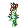
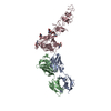
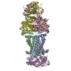
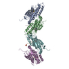
| Title | Crystal structure of yeast RNase III (Rnt1p) complexed with the product of dsRNA processing | ||||||
 Components Components |
| ||||||
 Keywords Keywords | HYDROLASE/RNA / RNase:RNA complex / ribonuclease III domain / double-stranded RNA-binding domain / endoribonuclease / dsRNA-specific RNase / double-stranded RNA / HYDROLASE-RNA complex | ||||||
| Function / homology |  Function and homology information Function and homology informationbox C/D sno(s)RNA processing / box H/ACA sno(s)RNA processing / regulation of fungal-type cell wall organization / termination of RNA polymerase II transcription, exosome-dependent / U1 snRNA 3'-end processing / U5 snRNA 3'-end processing / U4 snRNA 3'-end processing / ribonuclease III / ribonuclease III activity / termination of RNA polymerase II transcription ...box C/D sno(s)RNA processing / box H/ACA sno(s)RNA processing / regulation of fungal-type cell wall organization / termination of RNA polymerase II transcription, exosome-dependent / U1 snRNA 3'-end processing / U5 snRNA 3'-end processing / U4 snRNA 3'-end processing / ribonuclease III / ribonuclease III activity / termination of RNA polymerase II transcription / rRNA transcription / rRNA processing / double-stranded RNA binding / chromatin organization / nucleolus / nucleoplasm / nucleus Similarity search - Function | ||||||
| Biological species |  | ||||||
| Method |  X-RAY DIFFRACTION / SYNCHROTRON / MOLECULAR REPLACEMENT / Resolution: 2.5 Å X-RAY DIFFRACTION / SYNCHROTRON / MOLECULAR REPLACEMENT / Resolution: 2.5 Å | ||||||
 Authors Authors | Liang, Y.-H. / Ji, X. | ||||||
 Citation Citation | Journal: Mol.Cell / Year: 2014 Title: Structure of a Eukaryotic RNase III Postcleavage Complex Reveals a Double-Ruler Mechanism for Substrate Selection. Authors: Liang, Y.H. / Lavoie, M. / Comeau, M.A. / Abou Elela, S. / Ji, X. | ||||||
| History |
|
- Structure visualization
| Structure viewer | Molecule: MolmilJmol/JSmol |
|---|
- Downloads & links
-Download
| PDBx/mmCIF format | 4oog.cif.gz | 136.2 KB | Display | PDBx/mmCIF format |
|---|---|---|---|---|
| PDB format | pdb4oog.ent.gz | 101.7 KB | Display | PDB format |
| PDBx/mmJSON format | 4oog.json.gz | Tree view | PDBx/mmJSON format | |
| Others |  Other downloads Other downloads |
-Validation report
| Arichive directory | https://data.pdbj.org/pub/pdb/validation_reports/oo/4oogftp://data.pdbj.org/pub/pdb/validation_reports/oo/4oog | HTTPS FTP |
|---|
-Related structure data
| Related structure data |  1o0wS  1t4oS  1yywS  2nugS  3c4tS  3n3wS  3o2rS  3rv0S 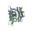 3rv1S S: Starting model for refinement |
|---|---|
| Similar structure data |


-Links
 PDBj
PDBj


- Assembly
| Deposited unit | 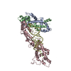
| ||||||||
|---|---|---|---|---|---|---|---|---|---|
| 1 | 
| ||||||||
| Unit cell |
|
-Components
| #1: Protein | Mass: 12804.515 Da / Num. of mol.: 2 / Fragment: N-terminal domain (UNP residues 42-151) Source method: isolated from a genetically manipulated source Source: (gene. exp.) Strain: ATCC 204508 / S288c / Gene: RNT1, YM9408.01C, YM9959.21, YMR239C / Plasmid: pQE31 / Production host:  #2: Protein | | Mass: 29934.600 Da / Num. of mol.: 1 Fragment: endonuclease domain and double-stranded RNA binding domain (UNP residues 197-457) Source method: isolated from a genetically manipulated source Source: (gene. exp.) Strain: ATCC 204508 / S288c / Gene: RNT1, YM9408.01C, YM9959.21, YMR239C / Plasmid: pQE31 / Production host: #3: RNA chain | | Mass: 10908.486 Da / Num. of mol.: 1 / Source method: obtained synthetically / Details: Derived from U5 snRNA 3' end cleavage product #4: Chemical | ChemComp-MG /   Mass: 24.305 Da / Num. of mol.: 4 / Source method: obtained synthetically / Formula: Mg Mass: 24.305 Da / Num. of mol.: 4 / Source method: obtained synthetically / Formula: Mg#5: Water | ChemComp-HOH / |  Mass: 18.015 Da / Num. of mol.: 98 / Source method: isolated from a natural source / Formula: H2O Mass: 18.015 Da / Num. of mol.: 98 / Source method: isolated from a natural source / Formula: H2O |
|---|
-Experimental details
-Experiment
| Experiment | Method: X-RAY DIFFRACTION / Number of used crystals: 1 |
|---|
- Sample preparation
| Crystal | Density Matthews: 3.35 Å3/Da / Density % sol: 63.26 % |
|---|---|
| Crystal grow | Temperature: 293 K / Method: vapor diffusion, hanging drop / pH: 8.5 Details: 25% PEG1000, 0.1 M Tris-HCl, pH 8.5, VAPOR DIFFUSION, HANGING DROP, temperature 293K |
-Data collection
| Diffraction | Mean temperature: 100 K | ||||||||||||||||||||||||||||||||||||||||||||||||||||||||||||||||||||||||||||||||
|---|---|---|---|---|---|---|---|---|---|---|---|---|---|---|---|---|---|---|---|---|---|---|---|---|---|---|---|---|---|---|---|---|---|---|---|---|---|---|---|---|---|---|---|---|---|---|---|---|---|---|---|---|---|---|---|---|---|---|---|---|---|---|---|---|---|---|---|---|---|---|---|---|---|---|---|---|---|---|---|---|---|
| Diffraction source | Source: SYNCHROTRON / Site: APS  / Beamline: 22-ID / Wavelength: 1 Å / Beamline: 22-ID / Wavelength: 1 Å | ||||||||||||||||||||||||||||||||||||||||||||||||||||||||||||||||||||||||||||||||
| Detector | Type: MARMOSAIC 300 mm CCD / Detector: CCD / Date: Nov 2, 2011 / Details: mirrors | ||||||||||||||||||||||||||||||||||||||||||||||||||||||||||||||||||||||||||||||||
| Radiation | Monochromator: double crystal Si(111) / Protocol: SINGLE WAVELENGTH / Monochromatic (M) / Laue (L): M / Scattering type: x-ray | ||||||||||||||||||||||||||||||||||||||||||||||||||||||||||||||||||||||||||||||||
| Radiation wavelength | Wavelength: 1 Å / Relative weight: 1 | ||||||||||||||||||||||||||||||||||||||||||||||||||||||||||||||||||||||||||||||||
| Reflection | Resolution: 2.5→40 Å / Num. obs: 31418 / % possible obs: 98.7 % / Observed criterion σ(I): -3 / Redundancy: 4.7 % / Biso Wilson estimate: 50.22 Å2 / Rmerge(I) obs: 0.066 / Net I/σ(I): 14.45 | ||||||||||||||||||||||||||||||||||||||||||||||||||||||||||||||||||||||||||||||||
| Reflection shell | Diffraction-ID: 1
|
- Processing
| Software |
| ||||||||||||||||||||||||||||||||||||||||||||||||||||||||||||||||
|---|---|---|---|---|---|---|---|---|---|---|---|---|---|---|---|---|---|---|---|---|---|---|---|---|---|---|---|---|---|---|---|---|---|---|---|---|---|---|---|---|---|---|---|---|---|---|---|---|---|---|---|---|---|---|---|---|---|---|---|---|---|---|---|---|---|
| Refinement | Method to determine structure: MOLECULAR REPLACEMENT Starting model: PDB ENTRIES 3RV0, 3RV1, 1YYW, 2NUG, 1O0W, 3N3W, 3O2R, 3C4T, AND 1T4O Resolution: 2.5→39.719 Å / Occupancy max: 1 / Occupancy min: 0.5 / SU ML: 0.33 / Cross valid method: THROUGHOUT / σ(F): 1.99 / Phase error: 33.23 / Stereochemistry target values: Engh & Huber
| ||||||||||||||||||||||||||||||||||||||||||||||||||||||||||||||||
| Solvent computation | Shrinkage radii: 0.98 Å / VDW probe radii: 1.2 Å / Solvent model: FLAT BULK SOLVENT MODEL / Bsol: 62.64 Å2 / ksol: 0.338 e/Å3 | ||||||||||||||||||||||||||||||||||||||||||||||||||||||||||||||||
| Displacement parameters | Biso max: 193.34 Å2 / Biso mean: 78.4156 Å2 / Biso min: 32.74 Å2
| ||||||||||||||||||||||||||||||||||||||||||||||||||||||||||||||||
| Refinement step | Cycle: LAST / Resolution: 2.5→39.719 Å
| ||||||||||||||||||||||||||||||||||||||||||||||||||||||||||||||||
| Refine LS restraints |
| ||||||||||||||||||||||||||||||||||||||||||||||||||||||||||||||||
| LS refinement shell | Refine-ID: X-RAY DIFFRACTION / Total num. of bins used: 7
|
