 Movie
Movie Controller

[English] 日本語
 Yorodumi
- PDB-3o2r: Structural flexibility in region involved in dimer formation of n... -
+ Open data

- Basic information
| Entry | Database: PDB / ID: 3o2r | ||||||
|---|---|---|---|---|---|---|---|
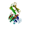
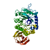
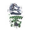
| Title | Structural flexibility in region involved in dimer formation of nuclease domain of Ribonuclase III (rnc) from Campylobacter jejuni | ||||||
 Components Components | (Ribonuclease III) x 2 | ||||||
 Keywords Keywords | HYDROLASE / Structural Genomics / Center for Structural Genomics of Infectious Diseases / CSGID / nuclease domain / ribonuclase III | ||||||
| Function / homology |  Function and homology information Function and homology informationribonuclease III / ribonuclease III activity / tRNA processing / mRNA processing / rRNA processing / double-stranded RNA binding / regulation of gene expression / rRNA binding / metal ion binding / cytoplasm Similarity search - Function | ||||||
| Biological species |  Campylobacter jejuni subsp. jejuni (Campylobacter) Campylobacter jejuni subsp. jejuni (Campylobacter) | ||||||
| Method |  X-RAY DIFFRACTION / SYNCHROTRON / MOLECULAR REPLACEMENT / Resolution: 1.251 Å X-RAY DIFFRACTION / SYNCHROTRON / MOLECULAR REPLACEMENT / Resolution: 1.251 Å | ||||||
 Authors Authors | Minasov, G. / Halavaty, A. / Shuvalova, L. / Dubrovska, I. / Winsor, J. / Papazisi, L. / Anderson, W.F. / Center for Structural Genomics of Infectious Diseases (CSGID) | ||||||
 Citation Citation | Journal: TO BE PUBLISHED Title: Structural Flexibility in Region Involved in Dimer Formation of Nuclease Domain of Ribonuclase III (rnc) from Campylobacter jejuni. Authors: Minasov, G. / Halavaty, A. / Shuvalova, L. / Dubrovska, I. / Winsor, J. / Papazisi, L. / Anderson, W.F. / Center for Structural Genomics of Infectious Diseases (CSGID) | ||||||
| History |
|
- Structure visualization
| Structure viewer | Molecule: MolmilJmol/JSmol |
|---|
- Downloads & links
-Download
| PDBx/mmCIF format | 3o2r.cif.gz | 296.9 KB | Display | PDBx/mmCIF format |
|---|---|---|---|---|
| PDB format | pdb3o2r.ent.gz | 242.6 KB | Display | PDB format |
| PDBx/mmJSON format | 3o2r.json.gz | Tree view | PDBx/mmJSON format | |
| Others |  Other downloads Other downloads |
-Validation report
| Arichive directory | https://data.pdbj.org/pub/pdb/validation_reports/o2/3o2rftp://data.pdbj.org/pub/pdb/validation_reports/o2/3o2r | HTTPS FTP |
|---|
-Related structure data
| Related structure data | 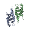 3n3wS S: Starting model for refinement |
|---|---|
| Similar structure data | |
| Other databases |


-Links
 PDBj
PDBj

- Assembly
| Deposited unit | 
| ||||||||
|---|---|---|---|---|---|---|---|---|---|
| 1 | 
| ||||||||
| 2 | 
| ||||||||
| Unit cell |
|
-Components
| #1: Protein | Mass: 19136.787 Da / Num. of mol.: 2 Source method: isolated from a genetically manipulated source Source: (gene. exp.) Campylobacter jejuni subsp. jejuni (Campylobacter)Strain: NCTC 11168 / Gene: CJSA_1547, rnc / Plasmid: pMCSG7 / Production host: References: UniProt: D3FPA5, UniProt: Q9PM40*PLUS, ribonuclease III #2: Protein | Mass: 19149.807 Da / Num. of mol.: 2 Source method: isolated from a genetically manipulated source Source: (gene. exp.) Campylobacter jejuni subsp. jejuni (Campylobacter)Strain: NCTC 11168 / Gene: CJSA_1547, rnc / Plasmid: pMCSG7 / Production host: References: UniProt: D3FPA5, UniProt: Q9PM40*PLUS, ribonuclease III #3: Chemical | ChemComp-CL /   Mass: 35.453 Da / Num. of mol.: 4 / Source method: obtained synthetically / Formula: Cl Mass: 35.453 Da / Num. of mol.: 4 / Source method: obtained synthetically / Formula: Cl#4: Water | ChemComp-HOH / |  Mass: 18.015 Da / Num. of mol.: 973 / Source method: isolated from a natural source / Formula: H2O Mass: 18.015 Da / Num. of mol.: 973 / Source method: isolated from a natural source / Formula: H2OSequence details | IN CHAINS B AND D, RESIDUE 31 DISPLAYS MICROHETEROGENEITY AND APPEARS AS BOTH A METHYLATED LYSINE ...IN CHAINS B AND D, RESIDUE 31 DISPLAYS MICROHETER | |
|---|
-Experimental details
-Experiment
| Experiment | Method: X-RAY DIFFRACTION / Number of used crystals: 1 |
|---|
- Sample preparation
| Crystal | Density Matthews: 1.83 Å3/Da / Density % sol: 32.6 % |
|---|---|
| Crystal grow | Temperature: 295 K / Method: vapor diffusion, sitting drop / pH: 4 Details: Protein solution: 7.6 mg/mL, 0.5M Sodium chloride, 0.01M Tris pH 8.3; Screen solution: PACT (B1), 0.1M MIB buffer pH 4.0, 25% w/v PEG1500, VAPOR DIFFUSION, SITTING DROP, temperature 295K |
-Data collection
| Diffraction | Mean temperature: 100 K |
|---|---|
| Diffraction source | Source: SYNCHROTRON / Site: APS  / Beamline: 21-ID-G / Wavelength: 0.97856 Å / Beamline: 21-ID-G / Wavelength: 0.97856 Å |
| Detector | Type: MARMOSAIC 300 mm CCD / Detector: CCD / Date: Jun 3, 2010 / Details: Beryllium lenses |
| Radiation | Monochromator: Diamond / Protocol: SINGLE WAVELENGTH / Monochromatic (M) / Laue (L): M / Scattering type: x-ray |
| Radiation wavelength | Wavelength: 0.97856 Å / Relative weight: 1 |
| Reflection | Resolution: 1.25→30 Å / Num. all: 151202 / Num. obs: 151202 / % possible obs: 99.6 % / Observed criterion σ(I): -3 / Redundancy: 3 % / Biso Wilson estimate: 11 Å2 / Rmerge(I) obs: 0.062 / Net I/σ(I): 16.4 |
| Reflection shell | Resolution: 1.25→1.27 Å / Redundancy: 2.4 % / Rmerge(I) obs: 0.359 / Mean I/σ(I) obs: 2.6 / Num. unique all: 7499 / % possible all: 99.4 |
- Processing
| Software |
| ||||||||||||||||||||||||||||||||||||||||||||||||||||||||||||||||||||||||||||||||||||||||||
|---|---|---|---|---|---|---|---|---|---|---|---|---|---|---|---|---|---|---|---|---|---|---|---|---|---|---|---|---|---|---|---|---|---|---|---|---|---|---|---|---|---|---|---|---|---|---|---|---|---|---|---|---|---|---|---|---|---|---|---|---|---|---|---|---|---|---|---|---|---|---|---|---|---|---|---|---|---|---|---|---|---|---|---|---|---|---|---|---|---|---|---|
| Refinement | Method to determine structure: MOLECULAR REPLACEMENT Starting model: PDB ENTRY 3N3W Resolution: 1.251→29.78 Å / Cor.coef. Fo:Fc: 0.977 / Cor.coef. Fo:Fc free: 0.966 / SU B: 0.866 / SU ML: 0.018 / Isotropic thermal model: Isotropic Individually refined / Cross valid method: THROUGHOUT / ESU R: 0.045 / ESU R Free: 0.045 / Stereochemistry target values: MAXIMUM LIKELIHOOD / Details: HYDROGENS HAVE BEEN ADDED IN THE RIDING POSITIONS
| ||||||||||||||||||||||||||||||||||||||||||||||||||||||||||||||||||||||||||||||||||||||||||
| Solvent computation | Ion probe radii: 0.8 Å / Shrinkage radii: 0.8 Å / VDW probe radii: 1.2 Å / Solvent model: BABINET MODEL WITH MASK | ||||||||||||||||||||||||||||||||||||||||||||||||||||||||||||||||||||||||||||||||||||||||||
| Displacement parameters | Biso mean: 11.409 Å2
| ||||||||||||||||||||||||||||||||||||||||||||||||||||||||||||||||||||||||||||||||||||||||||
| Refinement step | Cycle: LAST / Resolution: 1.251→29.78 Å
| ||||||||||||||||||||||||||||||||||||||||||||||||||||||||||||||||||||||||||||||||||||||||||
| Refine LS restraints |
| ||||||||||||||||||||||||||||||||||||||||||||||||||||||||||||||||||||||||||||||||||||||||||
| LS refinement shell | Resolution: 1.251→1.283 Å / Total num. of bins used: 20
|
