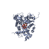
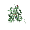
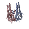
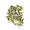
- PDB-3rv1: Crystal structure of the N-terminal and RNase III domains of K. p... -
+
Open data
ID or keywords:
Loading...
-
Basic information
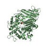
Entry
Database: PDB / ID: 3rv1
Title
Crystal structure of the N-terminal and RNase III domains of K. polysporus Dcr1 E224Q mutant
Components
K. polysporus Dcr1
Keywords
RNA BINDING PROTEIN / RNase III enzyme
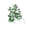
Function / homology
Function and homology information
U4 snRNA 3'-end processing / ribonuclease III activity / termination of RNA polymerase II transcription / rRNA processing / RNA binding / nucleoplasm / metal ion binding / identical protein binding Similarity search - Function
Substrate Binding Domain Of Dnak; Chain:A; Domain 2 - #260 / : / : / Dicers, N-terminal domain / Ribonuclease III domain / Ribonuclease iii, N-terminal Endonuclease Domain; Chain A / Ribonuclease III family signature. / Ribonuclease III domain / Ribonuclease III family domain profile. / Ribonuclease III family ...Substrate Binding Domain Of Dnak; Chain:A; Domain 2 - #260 / : / : / Dicers, N-terminal domain / Ribonuclease III domain / Ribonuclease iii, N-terminal Endonuclease Domain; Chain A / Ribonuclease III family signature. / Ribonuclease III domain / Ribonuclease III family domain profile. / Ribonuclease III family / Ribonuclease III domain / Double-stranded RNA binding motif / Double-stranded RNA binding motif / Substrate Binding Domain Of Dnak; Chain:A; Domain 2 / Ribonuclease III, endonuclease domain superfamily / Double stranded RNA-binding domain (dsRBD) profile. / Double-stranded RNA-binding domain / Up-down Bundle / Orthogonal Bundle / Mainly Alpha Similarity search - Domain/homology
In the structure databanks used in Yorodumi, some data are registered as the other names, "COVID-19 virus" and "2019-nCoV". Here are the details of the virus and the list of structure data.
Jan 31, 2019. EMDB accession codes are about to change! (news from PDBe EMDB page)
EMDB accession codes are about to change! (news from PDBe EMDB page)
The allocation of 4 digits for EMDB accession codes will soon come to an end. Whilst these codes will remain in use, new EMDB accession codes will include an additional digit and will expand incrementally as the available range of codes is exhausted. The current 4-digit format prefixed with “EMD-” (i.e. EMD-XXXX) will advance to a 5-digit format (i.e. EMD-XXXXX), and so on. It is currently estimated that the 4-digit codes will be depleted around Spring 2019, at which point the 5-digit format will come into force.
The EM Navigator/Yorodumi systems omit the EMD- prefix.
Related info.:Q: What is EMD? / ID/Accession-code notation in Yorodumi/EM Navigator
Yorodumi is a browser for structure data from EMDB, PDB, SASBDB, etc.
This page is also the successor to EM Navigator detail page, and also detail information page/front-end page for Omokage search.
The word "yorodu" (or yorozu) is an old Japanese word meaning "ten thousand". "mi" (miru) is to see.
Related info.:EMDB / PDB / SASBDB / Comparison of 3 databanks / Yorodumi Search / Aug 31, 2016. New EM Navigator & Yorodumi / Yorodumi Papers / Jmol/JSmol / Function and homology information / Changes in new EM Navigator and Yorodumi
 Movie
Movie Controller

 Yorodumi
 Open data
 Basic information
 Components
 Keywords
 Function and homology information
 Vanderwaltozyma polyspora (fungus)
 X-RAY DIFFRACTION /
  Authors
 Citation
 Structure visualization
 Downloads & links
 Other downloads


 PDBj
PDBj

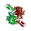
 Assembly
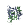

 Mass: 18.015 Da / Num. of mol.: 262 / Source method: isolated from a natural source / Formula: H2O
 Sample preparation
 / Beamline: 24-ID-E / Wavelength: 0.9792 Å
 Processing