 Movie
Movie Controller

[English] 日本語
 Yorodumi
- PDB-1l8w: Crystal Structure of Lyme Disease Variable Surface Antigen VlsE o... -
+ Open data

- Basic information
| Entry | Database: PDB / ID: 1l8w | ||||||
|---|---|---|---|---|---|---|---|
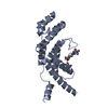
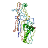
| Title | Crystal Structure of Lyme Disease Variable Surface Antigen VlsE of Borrelia burgdorferi | ||||||
 Components Components | VlsE1 | ||||||
 Keywords Keywords | IMMUNE SYSTEM / Variable Surface Protein / VMP-like sequence | ||||||
| Function / homology | Borrelia lipoprotein / Borrelia lipoprotein / cell outer membrane / Variable large protein Function and homology information Function and homology information | ||||||
| Biological species |  Borreliella burgdorferi (Lyme disease spirochete) Borreliella burgdorferi (Lyme disease spirochete) | ||||||
| Method |  X-RAY DIFFRACTION / SYNCHROTRON / MAD / Resolution: 2.3 Å X-RAY DIFFRACTION / SYNCHROTRON / MAD / Resolution: 2.3 Å | ||||||
 Authors Authors | Eicken, C. / Sharma, V. / Klabunde, T. / Lawrenz, M.B. / Hardham, J.M. / Norris, S.J. / Sacchettini, J.C. | ||||||
 Citation Citation | Journal: J.Biol.Chem. / Year: 2002 Title: Crystal structure of Lyme disease variable surface antigen VlsE of Borrelia burgdorferi. Authors: Eicken, C. / Sharma, V. / Klabunde, T. / Lawrenz, M.B. / Hardham, J.M. / Norris, S.J. / Sacchettini, J.C. | ||||||
| History |
| ||||||
| Remark 300 | BIOMOLECULE: 1, 2, 3, 4 THIS ENTRY CONTAINS THE CRYSTALLOGRAPHIC ASYMMETRIC UNIT WHICH CONSISTS OF ...BIOMOLECULE: 1, 2, 3, 4 THIS ENTRY CONTAINS THE CRYSTALLOGRAPHIC ASYMMETRIC UNIT WHICH CONSISTS OF 4 CHAIN(S). SEE REMARK 350 FOR INFORMATION ON GENERATING THE BIOLOGICAL MOLECULE(S). THE BIOLOGICAL ASSEMBLY IS ASSUMED TO BE A MONOMER. THE ASYMMETRIC UNIT CONTAINS 4 MONOMERS. |
- Structure visualization
| Structure viewer | Molecule: MolmilJmol/JSmol |
|---|
- Downloads & links
-Download
| PDBx/mmCIF format | 1l8w.cif.gz | 231.7 KB | Display | PDBx/mmCIF format |
|---|---|---|---|---|
| PDB format | pdb1l8w.ent.gz | 184.2 KB | Display | PDB format |
| PDBx/mmJSON format | 1l8w.json.gz | Tree view | PDBx/mmJSON format | |
| Others |  Other downloads Other downloads |
-Validation report
| Arichive directory | https://data.pdbj.org/pub/pdb/validation_reports/l8/1l8wftp://data.pdbj.org/pub/pdb/validation_reports/l8/1l8w | HTTPS FTP |
|---|
-Related structure data
| Similar structure data |
|---|


-Links
 PDBj
PDBj- Assembly
| Deposited unit | 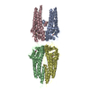
| ||||||||||||||||||||||||
|---|---|---|---|---|---|---|---|---|---|---|---|---|---|---|---|---|---|---|---|---|---|---|---|---|---|
| 1 | 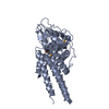
| ||||||||||||||||||||||||
| 2 | 
| ||||||||||||||||||||||||
| 3 | 
| ||||||||||||||||||||||||
| 4 | 
| ||||||||||||||||||||||||
| 5 | 
| ||||||||||||||||||||||||
| 6 | 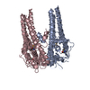
| ||||||||||||||||||||||||
| Unit cell |
| ||||||||||||||||||||||||
| Components on special symmetry positions |
| ||||||||||||||||||||||||
| Details | The biological assembly is assumed to be a monomer. The asymmetric unit contains 4 monomers. |
-Components
| #1: Protein | Mass: 35274.266 Da / Num. of mol.: 4 Source method: isolated from a genetically manipulated source Source: (gene. exp.) Borreliella burgdorferi (Lyme disease spirochete)Genus: Borrelia / Gene: vlsE / Production host: #2: Water | ChemComp-HOH / |  Mass: 18.015 Da / Num. of mol.: 1010 / Source method: isolated from a natural source / Formula: H2O Mass: 18.015 Da / Num. of mol.: 1010 / Source method: isolated from a natural source / Formula: H2OHas protein modification | Y | |
|---|
-Experimental details
-Experiment
| Experiment | Method: X-RAY DIFFRACTION / Number of used crystals: 1 |
|---|
- Sample preparation
| Crystal | Density Matthews: 2.01 Å3/Da / Density % sol: 38.72 % / Description: final refinement against 0.9797 data | ||||||||||||||||||||||||
|---|---|---|---|---|---|---|---|---|---|---|---|---|---|---|---|---|---|---|---|---|---|---|---|---|---|
| Crystal grow | Temperature: 291 K / Method: vapor diffusion, hanging drop / pH: 7 Details: PEG 1500, Hepes, pH 7.0, VAPOR DIFFUSION, HANGING DROP, temperature 291K | ||||||||||||||||||||||||
| Crystal grow | *PLUS pH: 8 / Method: batch method | ||||||||||||||||||||||||
| Components of the solutions | *PLUS
|
-Data collection
| Diffraction | Mean temperature: 100 K | ||||||||||||
|---|---|---|---|---|---|---|---|---|---|---|---|---|---|
| Diffraction source | Source: SYNCHROTRON / Site: APS  / Beamline: 14-BM-D / Wavelength: 0.9611,0.9797,0.9800 / Beamline: 14-BM-D / Wavelength: 0.9611,0.9797,0.9800 | ||||||||||||
| Detector | Type: ADSC QUANTUM 4 / Detector: CCD / Date: Dec 3, 2000 | ||||||||||||
| Radiation | Monochromator: CARS-design Si(111) double-bounce / Protocol: MAD / Monochromatic (M) / Laue (L): M / Scattering type: x-ray | ||||||||||||
| Radiation wavelength |
| ||||||||||||
| Reflection | Resolution: 2.3→90 Å / Num. all: 90139 / Num. obs: 90139 / % possible obs: 92.4 % / Observed criterion σ(F): 0 / Observed criterion σ(I): 0.5 / Biso Wilson estimate: 23 Å2 / Rsym value: 0.057 | ||||||||||||
| Reflection shell | Resolution: 2.3→2.38 Å / Rsym value: 0.138 / % possible all: 75 | ||||||||||||
| Reflection | *PLUS Lowest resolution: 90 Å / Rmerge(I) obs: 0.051 | ||||||||||||
| Reflection shell | *PLUS Highest resolution: 2.3 Å / % possible obs: 75 % / Rmerge(I) obs: 0.138 |
- Processing
| Software |
| ||||||||||||||||||||||||||||||||||||
|---|---|---|---|---|---|---|---|---|---|---|---|---|---|---|---|---|---|---|---|---|---|---|---|---|---|---|---|---|---|---|---|---|---|---|---|---|---|
| Refinement | Method to determine structure: MAD / Resolution: 2.3→82.43 Å / Rfactor Rfree error: 0.004 / Data cutoff high absF: 186825.69 / Data cutoff low absF: 0 / Isotropic thermal model: RESTRAINED / Cross valid method: THROUGHOUT / σ(F): 0 / Stereochemistry target values: Engh & Huber
| ||||||||||||||||||||||||||||||||||||
| Solvent computation | Solvent model: FLAT MODEL / Bsol: 84.1577 Å2 / ksol: 0.363979 e/Å3 | ||||||||||||||||||||||||||||||||||||
| Displacement parameters | Biso mean: 41.3 Å2
| ||||||||||||||||||||||||||||||||||||
| Refine analyze |
| ||||||||||||||||||||||||||||||||||||
| Refinement step | Cycle: LAST / Resolution: 2.3→82.43 Å
| ||||||||||||||||||||||||||||||||||||
| Refine LS restraints |
| ||||||||||||||||||||||||||||||||||||
| LS refinement shell | Resolution: 2.3→2.44 Å / Rfactor Rfree error: 0.012 / Total num. of bins used: 6
| ||||||||||||||||||||||||||||||||||||
| Xplor file |
| ||||||||||||||||||||||||||||||||||||
| Refinement | *PLUS Num. reflection obs: 41131 / Num. reflection Rfree: 4597 / Rfactor obs: 0.204 / Rfactor Rfree: 0.282 / Rfactor Rwork: 0.204 | ||||||||||||||||||||||||||||||||||||
| Solvent computation | *PLUS | ||||||||||||||||||||||||||||||||||||
| Displacement parameters | *PLUS | ||||||||||||||||||||||||||||||||||||
| Refine LS restraints | *PLUS
| ||||||||||||||||||||||||||||||||||||
| LS refinement shell | *PLUS Rfactor Rfree: 0.319 / Rfactor Rwork: 0.257 / Rfactor obs: 0.257 |
