 Movie
Movie Controller


+ Open data

- Basic information
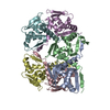
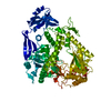
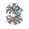
| Entry | Database: PDB / ID: 3bua | ||||||
|---|---|---|---|---|---|---|---|
| Title | Crystal Structure of TRF2 TRFH domain and APOLLO peptide complex | ||||||
 Components Components |
| ||||||
 Keywords Keywords | DNA BINDING PROTEIN / TRF2 TRFH domain Dimerization domain APOLLO peptide / Alternative splicing / Cell cycle / Chromosomal protein / DNA-binding / Nucleus / Phosphoprotein / Telomere / DNA damage / DNA repair / Polymorphism | ||||||
| Function / homology |  Function and homology information Function and homology informationaxonal transport of messenger ribonucleoprotein complex / negative regulation of telomere single strand break repair / telomeric 3' overhang formation / negative regulation of telomere maintenance via recombination / telomeric loop formation / negative regulation of telomere capping / negative regulation of telomere maintenance via semi-conservative replication / negative regulation of telomeric D-loop disassembly / protection from non-homologous end joining at telomere / RNA-templated DNA biosynthetic process ...axonal transport of messenger ribonucleoprotein complex / negative regulation of telomere single strand break repair / telomeric 3' overhang formation / negative regulation of telomere maintenance via recombination / telomeric loop formation / negative regulation of telomere capping / negative regulation of telomere maintenance via semi-conservative replication / negative regulation of telomeric D-loop disassembly / protection from non-homologous end joining at telomere / RNA-templated DNA biosynthetic process / negative regulation of telomere maintenance / negative regulation of t-circle formation / regulation of telomere maintenance via telomerase / telomere maintenance via telomere lengthening / telomeric D-loop disassembly / shelterin complex / Telomere C-strand synthesis initiation / double-stranded telomeric DNA binding / Telomere C-strand (Lagging Strand) Synthesis / G-rich strand telomeric DNA binding / nuclear telomere cap complex / telomere capping / Polymerase switching on the C-strand of the telomere / 5'-3' exonuclease activity / 5'-3' DNA exonuclease activity / Processive synthesis on the C-strand of the telomere / Removal of the Flap Intermediate from the C-strand / regulation of telomere maintenance / negative regulation of telomere maintenance via telomere lengthening / protein localization to chromosome, telomeric region / telomeric repeat DNA binding / negative regulation of cellular senescence / negative regulation of telomere maintenance via telomerase / positive regulation of telomere maintenance / Telomere Extension By Telomerase / interstrand cross-link repair / Packaging Of Telomere Ends / male germ cell nucleus / Recognition and association of DNA glycosylase with site containing an affected purine / Cleavage of the damaged purine / telomere maintenance / Recognition and association of DNA glycosylase with site containing an affected pyrimidine / Cleavage of the damaged pyrimidine / Inhibition of DNA recombination at telomere / Meiotic synapsis / Fanconi Anemia Pathway / beta-lactamase / beta-lactamase activity / DNA Damage/Telomere Stress Induced Senescence / double-strand break repair via nonhomologous end joining / cellular senescence / damaged DNA binding / Hydrolases; Acting on ester bonds / chromosome, telomeric region / nuclear body / axon / centrosome / protein-containing complex binding / enzyme binding / protein homodimerization activity / nucleoplasm / nucleus Similarity search - Function | ||||||
| Biological species |  Homo sapiens (human) Homo sapiens (human) | ||||||
| Method |  X-RAY DIFFRACTION / SYNCHROTRON / MOLECULAR REPLACEMENT / Resolution: 2.5 Å X-RAY DIFFRACTION / SYNCHROTRON / MOLECULAR REPLACEMENT / Resolution: 2.5 Å | ||||||
 Authors Authors | Chen, Y. / Yang, Y. / van Overbeek, M. / Donigian, J.R. / Baciu, P. / de Lange, T. / Lei, M. | ||||||
 Citation Citation | Journal: Science / Year: 2008 Title: A shared docking motif in TRF1 and TRF2 used for differential recruitment of telomeric proteins. Authors: Chen, Y. / Yang, Y. / van Overbeek, M. / Donigian, J.R. / Baciu, P. / de Lange, T. / Lei, M. | ||||||
| History |
|
- Structure visualization
| Structure viewer | Molecule: MolmilJmol/JSmol |
|---|
- Downloads & links
-Download
| PDBx/mmCIF format | 3bua.cif.gz | 182.1 KB | Display | PDBx/mmCIF format |
|---|---|---|---|---|
| PDB format | pdb3bua.ent.gz | 147.3 KB | Display | PDB format |
| PDBx/mmJSON format | 3bua.json.gz | Tree view | PDBx/mmJSON format | |
| Others |  Other downloads Other downloads |
-Validation report
| Arichive directory | https://data.pdbj.org/pub/pdb/validation_reports/bu/3buaftp://data.pdbj.org/pub/pdb/validation_reports/bu/3bua | HTTPS FTP |
|---|
-Related structure data
| Related structure data |  3bqoC 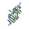 3bu8C 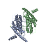 1h6pS C: citing same article ( S: Starting model for refinement |
|---|---|
| Similar structure data |


-Links
 PDBj
PDBj


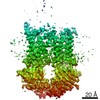
- Assembly
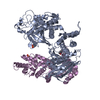
| Deposited unit | 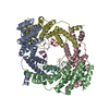
| ||||||||
|---|---|---|---|---|---|---|---|---|---|
| 1 |
| ||||||||
| 2 | 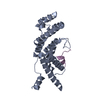
| ||||||||
| 3 | 
| ||||||||
| 4 | 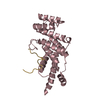
| ||||||||
| 5 | 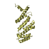
| ||||||||
| Unit cell |
|
-Components
| #1: Protein | Mass: 23724.646 Da / Num. of mol.: 4 / Fragment: TRFH domain, dimerization domain Source method: isolated from a genetically manipulated source Source: (gene. exp.) Homo sapiens (human) / Gene: TERF2, TRBF2, TRF2 / Plasmid: PET 28B-sumo / Species (production host): Escherichia coli / Production host:  #2: Protein/peptide | Mass: 4331.907 Da / Num. of mol.: 4 / Fragment: UNP residues 495-530 Source method: isolated from a genetically manipulated source Source: (gene. exp.) Homo sapiens (human) / Gene: DCLRE1B, SNM1B / Plasmid: PET 28B-sumo / Species (production host): Escherichia coli / Production host: #3: Water | ChemComp-HOH / |  Mass: 18.015 Da / Num. of mol.: 110 / Source method: isolated from a natural source / Formula: H2O Mass: 18.015 Da / Num. of mol.: 110 / Source method: isolated from a natural source / Formula: H2OSequence details | THE RESIDUE SER 495 IN CHAIN E,F,G,H CAME FROM A PROTEASE SPECIFIC DIGESTION. IT IS A RESIDUAL ...THE RESIDUE SER 495 IN CHAIN E,F,G,H CAME FROM A PROTEASE SPECIFIC DIGESTION. IT IS A RESIDUAL RESIDUE AFTER DIGESTION. | |
|---|
-Experimental details
-Experiment
| Experiment | Method: X-RAY DIFFRACTION |
|---|
- Sample preparation
| Crystal | Density Matthews: 2.03 Å3/Da / Density % sol: 39.53 % |
|---|---|
| Crystal grow | Temperature: 289 K / Method: vapor diffusion, hanging drop / pH: 5.6 Details: (NH4)2SO4 2.6 M DTT 1 mM MES 0.05 M pH 5.6 MgAc2 10 mM, VAPOR DIFFUSION, HANGING DROP, temperature 289K |
-Data collection
| Diffraction source | Source: SYNCHROTRON / Site: APS  / Beamline: 21-ID-D / Wavelength: 0.97869 Å / Beamline: 21-ID-D / Wavelength: 0.97869 Å |
|---|---|
| Detector | Type: MARMOSAIC 225 mm CCD / Detector: CCD / Date: Feb 8, 2007 |
| Radiation | Protocol: SINGLE WAVELENGTH / Monochromatic (M) / Laue (L): M / Scattering type: x-ray |
| Radiation wavelength | Wavelength: 0.97869 Å / Relative weight: 1 |
| Reflection | Resolution: 2.5→50 Å / Num. all: 31918 / Num. obs: 31885 / % possible obs: 99.9 % / Redundancy: 10.8 % / Biso Wilson estimate: 57.3 Å2 / Rmerge(I) obs: 0.086 / Net I/σ(I): 30 |
| Reflection shell | Resolution: 2.5→2.59 Å / Redundancy: 8 % / Rmerge(I) obs: 0.61 / Mean I/σ(I) obs: 2.51 / Num. unique all: 3110 / % possible all: 99.2 |
- Processing
| Software |
| ||||||||||||||||||||
|---|---|---|---|---|---|---|---|---|---|---|---|---|---|---|---|---|---|---|---|---|---|
| Refinement | Method to determine structure: MOLECULAR REPLACEMENT Starting model: 1H6P Resolution: 2.5→50 Å / σ(F): 0
| ||||||||||||||||||||
| Displacement parameters | Biso mean: 40.2 Å2 | ||||||||||||||||||||
| Refinement step | Cycle: LAST / Resolution: 2.5→50 Å
| ||||||||||||||||||||
| Refine LS restraints |
|
