 Movie
Movie Controller


+ Open data

- Basic information
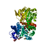
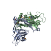
| Entry | Database: PDB / ID: 1h6p | ||||||
|---|---|---|---|---|---|---|---|
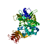
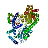
| Title | Dimeristion domain from human TRF2 | ||||||
 Components Components | TELOMERIC REPEAT BINDING FACTOR 2 | ||||||
 Keywords Keywords | TELOMERE BINDING / TRF2 / TELOMERE / DIMERISATION / TRFH / DNA-BINDING / NUCLEAR PROTEIN | ||||||
| Function / homology |  Function and homology information Function and homology informationaxonal transport of messenger ribonucleoprotein complex / negative regulation of telomere single strand break repair / negative regulation of telomere maintenance via recombination / telomeric loop formation / negative regulation of telomere capping / negative regulation of telomere maintenance via semi-conservative replication / negative regulation of telomeric D-loop disassembly / protection from non-homologous end joining at telomere / RNA-templated DNA biosynthetic process / negative regulation of telomere maintenance ...axonal transport of messenger ribonucleoprotein complex / negative regulation of telomere single strand break repair / negative regulation of telomere maintenance via recombination / telomeric loop formation / negative regulation of telomere capping / negative regulation of telomere maintenance via semi-conservative replication / negative regulation of telomeric D-loop disassembly / protection from non-homologous end joining at telomere / RNA-templated DNA biosynthetic process / negative regulation of telomere maintenance / negative regulation of t-circle formation / regulation of telomere maintenance via telomerase / telomeric D-loop disassembly / shelterin complex / Telomere C-strand synthesis initiation / double-stranded telomeric DNA binding / Telomere C-strand (Lagging Strand) Synthesis / G-rich strand telomeric DNA binding / nuclear telomere cap complex / telomere capping / Polymerase switching on the C-strand of the telomere / Processive synthesis on the C-strand of the telomere / Removal of the Flap Intermediate from the C-strand / regulation of telomere maintenance / negative regulation of telomere maintenance via telomere lengthening / protein localization to chromosome, telomeric region / telomeric repeat DNA binding / negative regulation of telomere maintenance via telomerase / positive regulation of telomere maintenance / negative regulation of cellular senescence / Telomere Extension By Telomerase / Packaging Of Telomere Ends / Recognition and association of DNA glycosylase with site containing an affected purine / Cleavage of the damaged purine / Recognition and association of DNA glycosylase with site containing an affected pyrimidine / Cleavage of the damaged pyrimidine / telomere maintenance / Inhibition of DNA recombination at telomere / Meiotic synapsis / male germ cell nucleus / DNA Damage/Telomere Stress Induced Senescence / cellular senescence / in utero embryonic development / chromosome, telomeric region / nuclear body / axon / protein-containing complex binding / enzyme binding / protein homodimerization activity / nucleoplasm / nucleus Similarity search - Function | ||||||
| Biological species |  HOMO SAPIENS (human) HOMO SAPIENS (human) | ||||||
| Method |  X-RAY DIFFRACTION / SYNCHROTRON / MIRAS / Resolution: 2.2 Å X-RAY DIFFRACTION / SYNCHROTRON / MIRAS / Resolution: 2.2 Å | ||||||
 Authors Authors | Chapman, L. / Fairall, L. / Rhodes, D. | ||||||
 Citation Citation | Journal: Mol.Cell / Year: 2001 Title: Structure of the Trfh Dimerization Domain of the Human Telomere Proteins Trf1 and Trf2 Authors: Fairall, L. / Chapman, L. / Moss, H. / de Lange, T. / Rhodes, D. #1: Journal: Nat.Genet. / Year: 1997 Title: Telomeric Localisation of Trf2, a Novel Human Telobox Protein Authors: Bilaud, T. / Brun, C. / Ancelin, K. / Koering, C.E. / Laroche, T. / Gilson, E. #2: Journal: Nat.Genet. / Year: 1997 Title: Human Telomere Contain Two Distinct Myb-Related Proteins, Trf1 and Trf2 Authors: Broccoli, D. / Smogorzewska, A. / Chong, L. / de Lange, T. | ||||||
| History |
|
- Structure visualization
| Structure viewer | Molecule: MolmilJmol/JSmol |
|---|
- Downloads & links
-Download
| PDBx/mmCIF format | 1h6p.cif.gz | 88.6 KB | Display | PDBx/mmCIF format |
|---|---|---|---|---|
| PDB format | pdb1h6p.ent.gz | 66.1 KB | Display | PDB format |
| PDBx/mmJSON format | 1h6p.json.gz | Tree view | PDBx/mmJSON format | |
| Others |  Other downloads Other downloads |
-Validation report
| Arichive directory | https://data.pdbj.org/pub/pdb/validation_reports/h6/1h6pftp://data.pdbj.org/pub/pdb/validation_reports/h6/1h6p | HTTPS FTP |
|---|
-Related structure data


-Links
 PDBj
PDBj


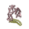
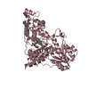
- Assembly
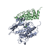
| Deposited unit | 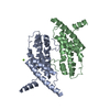
| ||||||||
|---|---|---|---|---|---|---|---|---|---|
| 1 |
| ||||||||
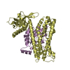
| Unit cell |
| ||||||||
| Components on special symmetry positions |
| ||||||||
| Noncrystallographic symmetry (NCS) | NCS oper: (Code: given Matrix: (-0.795, -0.0393, 0.6053), Vector: |
-Components
| #1: Protein | Mass: 23667.596 Da / Num. of mol.: 2 / Fragment: DIMERISATION DOMAIN RESIDUES 43-245 Source method: isolated from a genetically manipulated source Source: (gene. exp.) HOMO SAPIENS (human) / Cellular location: NUCLEUS / Plasmid: PET30A / Production host:  #2: Chemical | ChemComp-MG / |   Mass: 24.305 Da / Num. of mol.: 1 / Source method: obtained synthetically / Formula: Mg Mass: 24.305 Da / Num. of mol.: 1 / Source method: obtained synthetically / Formula: Mg#3: Water | ChemComp-HOH / |  Mass: 18.015 Da / Num. of mol.: 133 / Source method: isolated from a natural source / Formula: H2O Mass: 18.015 Da / Num. of mol.: 133 / Source method: isolated from a natural source / Formula: H2O |
|---|
-Experimental details
-Experiment
| Experiment | Method: X-RAY DIFFRACTION / Number of used crystals: 1 |
|---|
- Sample preparation
| Crystal | Density Matthews: 2.6 Å3/Da / Density % sol: 51.6 % | ||||||||||||||||||||||||||||||||||||||||||||||||||||||||||||
|---|---|---|---|---|---|---|---|---|---|---|---|---|---|---|---|---|---|---|---|---|---|---|---|---|---|---|---|---|---|---|---|---|---|---|---|---|---|---|---|---|---|---|---|---|---|---|---|---|---|---|---|---|---|---|---|---|---|---|---|---|---|
| Crystal grow | pH: 7 Details: PH7.0, 15-20% PEG 8000, 100-200 MM MAGNESIUM ACETATE, pH 7.00 | ||||||||||||||||||||||||||||||||||||||||||||||||||||||||||||
| Crystal grow | *PLUS pH: 8 / Method: vapor diffusion, hanging drop | ||||||||||||||||||||||||||||||||||||||||||||||||||||||||||||
| Components of the solutions | *PLUS
|
-Data collection
| Diffraction | Mean temperature: 100 K |
|---|---|
| Diffraction source | Source: SYNCHROTRON / Site: SRS  / Beamline: PX9.6 / Wavelength: 0.87 / Beamline: PX9.6 / Wavelength: 0.87 |
| Detector | Type: ADSC CCD / Detector: CCD / Date: Nov 15, 1999 / Details: MIRRORS |
| Radiation | Monochromator: SILICON / Protocol: SINGLE WAVELENGTH / Monochromatic (M) / Laue (L): M / Scattering type: x-ray |
| Radiation wavelength | Wavelength: 0.87 Å / Relative weight: 1 |
| Reflection | Resolution: 2.2→26.63 Å / Num. obs: 24415 / % possible obs: 99.1 % / Redundancy: 3.7 % / Biso Wilson estimate: 43.66 Å2 / Rmerge(I) obs: 0.052 / Rsym value: 0.052 / Net I/σ(I): 9.8 |
| Reflection shell | Resolution: 2.2→2.32 Å / Redundancy: 3.7 % / Rmerge(I) obs: 0.231 / Mean I/σ(I) obs: 3.1 / Rsym value: 0.231 / % possible all: 99.1 |
| Reflection | *PLUS Num. measured all: 89685 |
- Processing
| Software |
| ||||||||||||||||||||||||||||||||||||||||||||||||||||||||||||||||||||||||||||||||
|---|---|---|---|---|---|---|---|---|---|---|---|---|---|---|---|---|---|---|---|---|---|---|---|---|---|---|---|---|---|---|---|---|---|---|---|---|---|---|---|---|---|---|---|---|---|---|---|---|---|---|---|---|---|---|---|---|---|---|---|---|---|---|---|---|---|---|---|---|---|---|---|---|---|---|---|---|---|---|---|---|---|
| Refinement | Method to determine structure: MIRAS / Resolution: 2.2→23.72 Å / Rfactor Rfree error: 0.006 / Isotropic thermal model: RESTRAINED / Cross valid method: THROUGHOUT / σ(F): 2
| ||||||||||||||||||||||||||||||||||||||||||||||||||||||||||||||||||||||||||||||||
| Solvent computation | Solvent model: FLAT MODEL / ksol: 0.363168 e/Å3 | ||||||||||||||||||||||||||||||||||||||||||||||||||||||||||||||||||||||||||||||||
| Displacement parameters | Biso mean: 42.3 Å2
| ||||||||||||||||||||||||||||||||||||||||||||||||||||||||||||||||||||||||||||||||
| Refine analyze |
| ||||||||||||||||||||||||||||||||||||||||||||||||||||||||||||||||||||||||||||||||
| Refinement step | Cycle: LAST / Resolution: 2.2→23.72 Å
| ||||||||||||||||||||||||||||||||||||||||||||||||||||||||||||||||||||||||||||||||
| Refine LS restraints |
| ||||||||||||||||||||||||||||||||||||||||||||||||||||||||||||||||||||||||||||||||
| LS refinement shell | Resolution: 2.2→2.34 Å / Rfactor Rfree error: 0.016 / Total num. of bins used: 6
| ||||||||||||||||||||||||||||||||||||||||||||||||||||||||||||||||||||||||||||||||
| Xplor file |
| ||||||||||||||||||||||||||||||||||||||||||||||||||||||||||||||||||||||||||||||||
| Software | *PLUS Name: CNS / Version: 1 / Classification: refinement | ||||||||||||||||||||||||||||||||||||||||||||||||||||||||||||||||||||||||||||||||
| Refine LS restraints | *PLUS
| ||||||||||||||||||||||||||||||||||||||||||||||||||||||||||||||||||||||||||||||||
| LS refinement shell | *PLUS Rfactor Rwork: 0.24 |
