 Movie
Movie Controller


+ Open data

- Basic information
| Entry | Database: PDB / ID: 1blx | ||||||
|---|---|---|---|---|---|---|---|
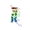
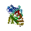
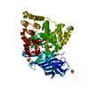
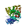
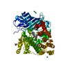
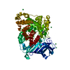
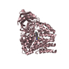
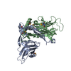
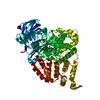
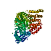
| Title | P19INK4D/CDK6 COMPLEX | ||||||
 Components Components |
| ||||||
 Keywords Keywords | COMPLEX (INHIBITOR PROTEIN/KINASE) / INHIBITOR PROTEIN / CYCLIN-DEPENDENT KINASE / CELL CYCLE CONTROL / ALPHA/BETA / COMPLEX (INHIBITOR PROTEIN-KINASE) / COMPLEX (INHIBITOR PROTEIN-KINASE) complex | ||||||
| Function / homology |  Function and homology information Function and homology informationcyclin D2-CDK6 complex / cyclin D3-CDK6 complex / cyclin D2-CDK4 complex / cyclin D1-CDK6 complex / cell dedifferentiation / Evasion of Oncogene Induced Senescence Due to Defective p16INK4A binding to CDK4 and CDK6 / Evasion of Oxidative Stress Induced Senescence Due to Defective p16INK4A binding to CDK4 and CDK6 / Drug-mediated inhibition of CDK4/CDK6 activity / FBXO family protein binding / lateral ventricle development ...cyclin D2-CDK6 complex / cyclin D3-CDK6 complex / cyclin D2-CDK4 complex / cyclin D1-CDK6 complex / cell dedifferentiation / Evasion of Oncogene Induced Senescence Due to Defective p16INK4A binding to CDK4 and CDK6 / Evasion of Oxidative Stress Induced Senescence Due to Defective p16INK4A binding to CDK4 and CDK6 / Drug-mediated inhibition of CDK4/CDK6 activity / FBXO family protein binding / lateral ventricle development / autophagic cell death / negative regulation of cell cycle G1/S phase transition / negative regulation of myeloid cell differentiation / type B pancreatic cell development / negative regulation of monocyte differentiation / regulation of cell motility / astrocyte development / cyclin-dependent protein serine/threonine kinase inhibitor activity / dentate gyrus development / negative regulation of intrinsic apoptotic signaling pathway in response to DNA damage / gliogenesis / Regulation of RUNX1 Expression and Activity / response to vitamin D / regulation of hematopoietic stem cell differentiation / positive regulation of cell-matrix adhesion / negative regulation of G1/S transition of mitotic cell cycle / generation of neurons / DNA synthesis involved in DNA repair / Defective binding of RB1 mutants to E2F1,(E2F2, E2F3) / negative regulation of cell cycle / negative regulation of cellular senescence / regulation of G1/S transition of mitotic cell cycle / negative regulation of cell differentiation / response to retinoic acid / cyclin-dependent kinase / hematopoietic stem cell differentiation / cyclin-dependent protein serine/threonine kinase activity / negative regulation of osteoblast differentiation / response to UV / cyclin-dependent protein kinase holoenzyme complex / regulation of G2/M transition of mitotic cell cycle / Notch signaling pathway / ruffle / cyclin binding / regulation of erythrocyte differentiation / sensory perception of sound / G1/S transition of mitotic cell cycle / negative regulation of cell growth / Oncogene Induced Senescence / positive regulation of fibroblast proliferation / negative regulation of epithelial cell proliferation / response to virus / Cyclin D associated events in G1 / T cell differentiation in thymus / regulation of gene expression / Senescence-Associated Secretory Phenotype (SASP) / Oxidative Stress Induced Senescence / regulation of cell cycle / negative regulation of cell population proliferation / protein serine kinase activity / cell division / DNA damage response / positive regulation of gene expression / centrosome / protein kinase binding / negative regulation of transcription by RNA polymerase II / signal transduction / nucleoplasm / ATP binding / nucleus / cytoplasm / cytosol Similarity search - Function | ||||||
| Biological species |  Homo sapiens (human) Homo sapiens (human) | ||||||
| Method |  X-RAY DIFFRACTION / SYNCHROTRON / MOLECULAR REPLACEMENT / Resolution: 1.9 Å X-RAY DIFFRACTION / SYNCHROTRON / MOLECULAR REPLACEMENT / Resolution: 1.9 Å | ||||||
 Authors Authors | Brotherton, D.H. / Dhanaraj, V. / Wick, S. / Brizuela, L. / Domaille, P.J. / Volyanik, E. / Xu, X. / Parisini, E. / Smith, B.O. / Archer, S.J. ...Brotherton, D.H. / Dhanaraj, V. / Wick, S. / Brizuela, L. / Domaille, P.J. / Volyanik, E. / Xu, X. / Parisini, E. / Smith, B.O. / Archer, S.J. / Serrano, M. / Brenner, S.L. / Blundell, T.L. / Laue, E.D. | ||||||
 Citation Citation | Journal: Nature / Year: 1998 Title: Crystal structure of the complex of the cyclin D-dependent kinase Cdk6 bound to the cell-cycle inhibitor p19INK4d. Authors: Brotherton, D.H. / Dhanaraj, V. / Wick, S. / Brizuela, L. / Domaille, P.J. / Volyanik, E. / Xu, X. / Parisini, E. / Smith, B.O. / Archer, S.J. / Serrano, M. / Brenner, S.L. / Blundell, T.L. / Laue, E.D. #1: Journal: Nature / Year: 1998Title: Erratum. Crystal Structure of the Complex of the Cyclin D-Dependent Kinase Cdk6 Bound to the Cell-Cycle Inhibitor P19Ink4D Authors: Brotherton, D.H. / Dhanaraj, V. / Wick, S. / Brizuela, L. / Domaille, P.J. / Volyanik, E. / Xu, X. / Parisini, E. / Smith, B.O. / Archer, S.J. / Serrano, M. / Brenner, S.L. / Blundell, T.L. / Laue, E.D. | ||||||
| History |
|
- Structure visualization
| Structure viewer | Molecule: MolmilJmol/JSmol |
|---|
- Downloads & links
-Download
| PDBx/mmCIF format | 1blx.cif.gz | 110.5 KB | Display | PDBx/mmCIF format |
|---|---|---|---|---|
| PDB format | pdb1blx.ent.gz | 83.9 KB | Display | PDB format |
| PDBx/mmJSON format | 1blx.json.gz | Tree view | PDBx/mmJSON format | |
| Others |  Other downloads Other downloads |
-Validation report
| Arichive directory | https://data.pdbj.org/pub/pdb/validation_reports/bl/1blxftp://data.pdbj.org/pub/pdb/validation_reports/bl/1blx | HTTPS FTP |
|---|
-Related structure data


-Links
 PDBj
PDBj


- Assembly
| Deposited unit | 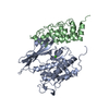
| ||||||||
|---|---|---|---|---|---|---|---|---|---|
| 1 |
| ||||||||
| Unit cell |
|
-Components
| #1: Protein | Mass: 36987.328 Da / Num. of mol.: 1 Source method: isolated from a genetically manipulated source Source: (gene. exp.) Homo sapiens (human) / Cell line: SF9 / Plasmid: GST FUSION PLASMID / Cell line (production host): SF9 / Production host:   Spodoptera frugiperda (fall armyworm) / Strain (production host): BL21 (DE3) / References: UniProt: Q00534 Spodoptera frugiperda (fall armyworm) / Strain (production host): BL21 (DE3) / References: UniProt: Q00534 | ||
|---|---|---|---|
| #2: Protein | Mass: 17832.375 Da / Num. of mol.: 1 Source method: isolated from a genetically manipulated source Source: (gene. exp.)  | ||
| #3: Chemical |   Mass: 40.078 Da / Num. of mol.: 1 / Source method: obtained synthetically / Formula: Ca Mass: 40.078 Da / Num. of mol.: 1 / Source method: obtained synthetically / Formula: Ca#4: Water | ChemComp-HOH / |  Mass: 18.015 Da / Num. of mol.: 294 / Source method: isolated from a natural source / Formula: H2O Mass: 18.015 Da / Num. of mol.: 294 / Source method: isolated from a natural source / Formula: H2O |
-Experimental details
-Experiment
| Experiment | Method: X-RAY DIFFRACTION / Number of used crystals: 1 |
|---|
- Sample preparation
| Crystal | Density Matthews: 2.42 Å3/Da / Density % sol: 49.27 % | ||||||||||||||||||||
|---|---|---|---|---|---|---|---|---|---|---|---|---|---|---|---|---|---|---|---|---|---|
| Crystal grow | pH: 7.5 / Details: pH 7.5 | ||||||||||||||||||||
| Crystal grow | *PLUS Temperature: 4 ℃ / pH: 6.5 / Method: vapor diffusion, hanging drop | ||||||||||||||||||||
| Components of the solutions | *PLUS
|
-Data collection
| Diffraction | Mean temperature: 279 K |
|---|---|
| Diffraction source | Source: SYNCHROTRON / Site: ESRF  / Beamline: ID14-3 / Wavelength: 0.9475 / Beamline: ID14-3 / Wavelength: 0.9475 |
| Detector | Type: MARRESEARCH / Detector: CCD / Date: Feb 1, 1998 / Details: MIRRORS |
| Radiation | Monochromator: SI(111) / Monochromatic (M) / Laue (L): M / Scattering type: x-ray |
| Radiation wavelength | Wavelength: 0.9475 Å / Relative weight: 1 |
| Reflection | Resolution: 1.9→18.54 Å / Num. obs: 41965 / % possible obs: 98.5 % / Redundancy: 4 % / Rmerge(I) obs: 0.054 / Rsym value: 0.054 / Net I/σ(I): 8.4 |
| Reflection shell | Resolution: 1.9→2 Å / Redundancy: 3.6 % / Rmerge(I) obs: 0.213 / Mean I/σ(I) obs: 1.9 / Rsym value: 0.327 / % possible all: 97.7 |
| Reflection | *PLUS Num. measured all: 166218 |
- Processing
| Software |
| ||||||||||||||||
|---|---|---|---|---|---|---|---|---|---|---|---|---|---|---|---|---|---|
| Refinement | Method to determine structure: MOLECULAR REPLACEMENT Starting model: PDB ENTRIES 1HCK AND 1AP7 Resolution: 1.9→18.54 Å / Cross valid method: FREE R / σ(F): 0
| ||||||||||||||||
| Refinement step | Cycle: LAST / Resolution: 1.9→18.54 Å
| ||||||||||||||||
| Software | *PLUS Name: REFMAC / Classification: refinement | ||||||||||||||||
| Refinement | *PLUS Rfactor obs: 0.2 | ||||||||||||||||
| Solvent computation | *PLUS | ||||||||||||||||
| Displacement parameters | *PLUS | ||||||||||||||||
| Refine LS restraints | *PLUS
|
