[English] 日本語
Yorodumi
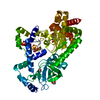
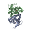
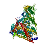
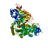
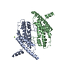
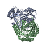
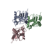
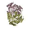
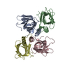
- PDB-4egw: The structure of the soluble domain of CorA from Methanocaldococc... -

+
Open data


ID or keywords:

Loading...

-
Basic information

Entry
Database: PDB / ID: 4egw
TitleThe structure of the soluble domain of CorA from Methanocaldococcus jannaschii
ComponentsMagnesium transport protein CorA
KeywordsMETAL TRANSPORT / magnesium transporter / magnesium binding / CORA
Function / homology
Function and homology information


cobalt ion transmembrane transporter activity / magnesium ion transmembrane transporter activity / cobalt ion binding / magnesium ion binding / identical protein binding / plasma membrane
Similarity search - Function
Magnesium transport protein CorA, transmembrane region / CorA soluble domain-like / Magnesium/cobalt transport protein CorA / CorA, cytoplasmic domain / CorA, transmembrane region / Mg2+ transporter protein, CorA-like/Zinc transport protein ZntB / CorA-like Mg2+ transporter protein / Beta Polymerase; domain 2 / Methane Monooxygenase Hydroxylase; Chain G, domain 1 / Up-down Bundle ...Magnesium transport protein CorA, transmembrane region / CorA soluble domain-like / Magnesium/cobalt transport protein CorA / CorA, cytoplasmic domain / CorA, transmembrane region / Mg2+ transporter protein, CorA-like/Zinc transport protein ZntB / CorA-like Mg2+ transporter protein / Beta Polymerase; domain 2 / Methane Monooxygenase Hydroxylase; Chain G, domain 1 / Up-down Bundle / 2-Layer Sandwich / Mainly Alpha / Alpha Beta
Similarity search - Domain/homology
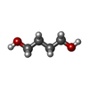
1,4-BUTANEDIOL / HEXANE-1,6-DIOL / S-1,2-PROPANEDIOL / Cobalt/magnesium transport protein CorA
Similarity search - Component
Biological speciesMethanocaldococcus jannaschii (archaea)
MethodX-RAY DIFFRACTION / SYNCHROTRON / SAD, molecular replacement / Resolution: 2.5 Å
AuthorsGuskov, A. / Nordin, N. / Reynaud, A. / Engman, H. / Lundback, A.-K. / Jong, A.J.O. / Cornvik, T. / Phua, T. / Eshaghi, S.
CitationJournal: Proc.Natl.Acad.Sci.USA / Year: 2012
Title: Structural insights into the mechanisms of Mg2+ uptake, transport, and gating by CorA
Authors: Guskov, A. / Nordin, N. / Reynaud, A. / Engman, H. / Lundback, A.K. / Jong, A.J. / Cornvik, T. / Phua, T. / Eshaghi, S.
History
DepositionApr 2, 2012Deposition site: RCSB / Processing site: PDBJ
Revision 1.0Oct 31, 2012Provider: repository / Type: Initial release
Revision 1.1Jul 24, 2013Group: Database references
Revision 1.2Mar 20, 2024Group: Data collection / Database references / Derived calculations
Category: chem_comp_atom / chem_comp_bond ...chem_comp_atom / chem_comp_bond / database_2 / struct_ref_seq_dif / struct_site
Item: _database_2.pdbx_DOI / _database_2.pdbx_database_accession ..._database_2.pdbx_DOI / _database_2.pdbx_database_accession / _struct_ref_seq_dif.details / _struct_site.pdbx_auth_asym_id / _struct_site.pdbx_auth_comp_id / _struct_site.pdbx_auth_seq_id

-
Structure visualization

Structure viewerMolecule:
MolmilJmol/JSmol

Downloads & links

-
Assembly

Deposited unit
A: Magnesium transport protein CorA
B: Magnesium transport protein CorA
hetero molecules


Theoretical massNumber of molelcules
Total (without water)68,70134
Polymers65,7902
Non-polymers2,91132
Water1,72996
1


  • Idetical with deposited unit
  • defined by author
TypeNameSymmetry operationNumber
identity operation1_555x,y,z1
Unit cell
Length a, b, c (Å)68.290, 68.290, 241.940
Angle α, β, γ (deg.)90.00, 90.00, 120.00
Int Tables number169
Space group name H-MP61

-
Components

-
Protein , 1 types, 2 molecules AB

#1: Protein Magnesium transport protein CorA


Mass: 32894.984 Da / Num. of mol.: 2 / Fragment: Soluble domain, UNP RESIDUES 1-258
Source method: isolated from a genetically manipulated source
Source: (gene. exp.) Methanocaldococcus jannaschii (archaea)
Strain: DSM 2661 / Gene: corA, MJ1033 / Production host: Escherichia coli (E. coli) / References: UniProt: Q58439

-
Non-polymers , 5 types, 128 molecules

#2: Chemical
ChemComp-HEZ / HEXANE-1,6-DIOL


Mass: 118.174 Da / Num. of mol.: 13 / Source method: obtained synthetically / Formula: C6H14O2
#3: Chemical
ChemComp-BU1 / 1,4-BUTANEDIOL


Mass: 90.121 Da / Num. of mol.: 6 / Source method: obtained synthetically / Formula: C4H10O2
#4: Chemical
ChemComp-PGO / S-1,2-PROPANEDIOL


Mass: 76.094 Da / Num. of mol.: 10 / Source method: obtained synthetically / Formula: C3H8O2
#5: Chemical ChemComp-MG / MAGNESIUM ION


Mass: 24.305 Da / Num. of mol.: 3 / Source method: obtained synthetically / Formula: Mg
#6: Water ChemComp-HOH / water


Mass: 18.015 Da / Num. of mol.: 96 / Source method: isolated from a natural source / Formula: H2O

-
Experimental details

-
Experiment

ExperimentMethod: X-RAY DIFFRACTION / Number of used crystals: 1

-
Sample preparation

CrystalDensity Matthews: 2.48 Å3/Da / Density % sol: 50.31 %
Crystal growTemperature: 298 K / Method: vapor diffusion, sitting drop / pH: 7.5
Details: 28% PEG 8000, HEPES-MOPS, Alcohol mix, pH 7.5, VAPOR DIFFUSION, SITTING DROP, temperature 298K

-
Data collection

DiffractionMean temperature: 100 K
Diffraction sourceSource: SYNCHROTRON / Site: NSRRC / Beamline: BL13C1 / Wavelength: 0.975 Å
DetectorType: RAYONIX MX300HE / Detector: CCD / Date: Apr 21, 2011
RadiationProtocol: SINGLE WAVELENGTH / Monochromatic (M) / Laue (L): M / Scattering type: x-ray
Radiation wavelengthWavelength: 0.975 Å / Relative weight: 1
Reflection twinOperator: k,h,-l / Fraction: 0.5
ReflectionResolution: 2.5→50 Å / Num. obs: 21977 / % possible obs: 99.8 % / Observed criterion σ(F): 0 / Observed criterion σ(I): 0
Reflection shellResolution: 2.5→2.61 Å / % possible all: 99.8

-
Processing

Software
NameVersionClassification
Auto-Rickshawphasing
PHENIX(phenix.refine: 1.7.1_743)refinement
XDSdata reduction
XDSdata scaling
RefinementMethod to determine structure: SAD, molecular replacement / Resolution: 2.5→47.692 Å / σ(F): 2.01 / Phase error: 20.45 / Stereochemistry target values: Engh & Huber
RfactorNum. reflection% reflectionSelection details
Rfree0.1949 1099 5 %RANDOM
Rwork0.153 ---
obs0.1531 21977 99.86 %-
Solvent computationShrinkage radii: 0.83 Å / VDW probe radii: 1.1 Å / Solvent model: FLAT BULK SOLVENT MODEL / Bsol: 74.293 Å2 / ksol: 0.37 e/Å3
Refinement stepCycle: LAST / Resolution: 2.5→47.692 Å
ProteinNucleic acidLigandSolventTotal
Num. atoms4124 0 193 96 4413
Refine LS restraints
Refine-IDTypeDev idealNumber
X-RAY DIFFRACTIONf_bond_d0.0074373
X-RAY DIFFRACTIONf_angle_d1.3435829
X-RAY DIFFRACTIONf_dihedral_angle_d21.4521722
X-RAY DIFFRACTIONf_chiral_restr0.084677
X-RAY DIFFRACTIONf_plane_restr0.005707
LS refinement shell

Refine-ID: X-RAY DIFFRACTION / Total num. of bins used: 8

Resolution (Å)Rfactor RfreeNum. reflection RfreeRfactor RworkNum. reflection Rwork% reflection obs (%)
2.5011-2.6150.26831380.2079261194
2.615-2.75280.22461370.1951259695
2.7528-2.92520.21891360.1855260395
2.9252-3.1510.211370.1663259095
3.151-3.4680.18891370.1505261695
3.468-3.96950.20691380.1345261695
3.9695-4.99990.15981370.119260595
4.9999-42.29210.20351390.1571262695
Refinement TLS params.

Method: refined / Refine-ID: X-RAY DIFFRACTION

IDL112)L122)L132)L222)L232)L332)S11 (Å °)S12 (Å °)S13 (Å °)S21 (Å °)S22 (Å °)S23 (Å °)S31 (Å °)S32 (Å °)S33 (Å °)T112)T122)T132)T222)T232)T332)Origin x (Å)Origin y (Å)Origin z (Å)
13.6006-1.4136-0.76363.10010.31673.21590.0070.1513-0.6322-0.32780.2435-0.12170.90580.2772-0.18930.5595-0.04630.01350.2764-0.19630.641720.7166-41.8525-12.6917
21.17382.20870.32685.01830.28122.0465-0.0882-0.2038-0.43520.06670.0270.10460.2484-0.13490.02580.2194-0.09370.09080.1734-0.04890.629123.0957-33.1505-2.3745
37.4089-2.88022.41093.88120.09644.03880.37151.4215-0.7678-1.1292-0.1931-0.15120.4250.8881-0.17220.8504-0.13060.16880.5643-0.18060.651229.3458-34.8714-20.9801
40.9958-0.2011-0.17013.20580.70671.5563-0.0763-0.14780.1717-0.09970.06980.0251-0.20640.02470.00920.1869-0.03320.02070.0856-0.16250.582529.1383-12.3774-8.3838
51.05391.192-0.55842.30520.75632.3076-0.089-0.25510.4702-0.36550.1582-0.3762-0.55810.66740.08350.4069-0.208-0.10640.53470.0230.756249.21782.4366-24.7521
61.70970.66230.31822.33160.11782.2473-0.29790.30860.1872-0.19360.1283-0.0416-0.45160.42890.10270.2986-0.13510.00140.3575-0.07770.689540.9277-2.5841-36.2326
72.18870.4115-0.59182.0256-0.06151.94870.0771-0.3494-0.05120.0685-0.1484-0.22980.111-0.03480.07560.1891-0.032-0.02930.2769-0.14020.476330.3153-14.5558-27.8229
82.49121.26991.73491.7341-0.16872.8682-0.01930.0149-0.4122-0.144-0.0769-0.2790.2783-0.33440.13270.2867-0.05970.030.3049-0.11570.57525.0696-22.9032-31.9525
Refinement TLS group
IDRefine-IDRefine TLS-IDSelection details
1X-RAY DIFFRACTION1CHAIN A AND (RESSEQ 1:37)
2X-RAY DIFFRACTION2CHAIN A AND (RESSEQ 38:109)
3X-RAY DIFFRACTION3CHAIN A AND (RESSEQ 110:133)
4X-RAY DIFFRACTION4CHAIN A AND (RESSEQ 134:250)
5X-RAY DIFFRACTION5CHAIN B AND (RESSEQ 1:28)
6X-RAY DIFFRACTION6CHAIN B AND (RESSEQ 29:109)
7X-RAY DIFFRACTION7CHAIN B AND (RESSEQ 110:210)
8X-RAY DIFFRACTION8CHAIN B AND (RESSEQ 211:250)

+
About Yorodumi

-
News

-
Feb 9, 2022. New format data for meta-information of EMDB entries

New format data for meta-information of EMDB entries

  • Version 3 of the EMDB header file is now the official format.
  • The previous official version 1.9 will be removed from the archive.

Related info.:EMDB header

External links:wwPDB to switch to version 3 of the EMDB data model

-
Aug 12, 2020. Covid-19 info

Covid-19 info

URL: https://pdbjlvh1.pdbj.org/emnavi/covid19.php

New page: Covid-19 featured information page in EM Navigator.

Related info.:Covid-19 info / Mar 5, 2020. Novel coronavirus structure data

+
Mar 5, 2020. Novel coronavirus structure data

Novel coronavirus structure data

Related info.:Yorodumi Speices / Aug 12, 2020. Covid-19 info

External links:COVID-19 featured content - PDBj / Molecule of the Month (242):Coronavirus Proteases

+
Jan 31, 2019. EMDB accession codes are about to change! (news from PDBe EMDB page)

EMDB accession codes are about to change! (news from PDBe EMDB page)

  • The allocation of 4 digits for EMDB accession codes will soon come to an end. Whilst these codes will remain in use, new EMDB accession codes will include an additional digit and will expand incrementally as the available range of codes is exhausted. The current 4-digit format prefixed with “EMD-” (i.e. EMD-XXXX) will advance to a 5-digit format (i.e. EMD-XXXXX), and so on. It is currently estimated that the 4-digit codes will be depleted around Spring 2019, at which point the 5-digit format will come into force.
  • The EM Navigator/Yorodumi systems omit the EMD- prefix.

Related info.:Q: What is EMD? / ID/Accession-code notation in Yorodumi/EM Navigator

External links:EMDB Accession Codes are Changing Soon! / Contact to PDBj

+
Jul 12, 2017. Major update of PDB

Major update of PDB

  • wwPDB released updated PDB data conforming to the new PDBx/mmCIF dictionary.
  • This is a major update changing the version number from 4 to 5, and with Remediation, in which all the entries are updated.
  • In this update, many items about electron microscopy experimental information are reorganized (e.g. em_software).
  • Now, EM Navigator and Yorodumi are based on the updated data.

External links:wwPDB Remediation / Enriched Model Files Conforming to OneDep Data Standards Now Available in the PDB FTP Archive

-
Yorodumi

Thousand views of thousand structures

  • Yorodumi is a browser for structure data from EMDB, PDB, SASBDB, etc.
  • This page is also the successor to EM Navigator detail page, and also detail information page/front-end page for Omokage search.
  • The word "yorodu" (or yorozu) is an old Japanese word meaning "ten thousand". "mi" (miru) is to see.

Related info.:EMDB / PDB / SASBDB / Comparison of 3 databanks / Yorodumi Search / Aug 31, 2016. New EM Navigator & Yorodumi / Yorodumi Papers / Jmol/JSmol / Function and homology information / Changes in new EM Navigator and Yorodumi

Read more