- PDB-2pqa: Crystal Structure of Full-length Human RPA 14/32 Heterodimer -
+
Open data
ID or keywords:
Loading...
-
Basic information
Entry
Database: PDB / ID: 2pqa
Title
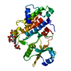
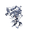
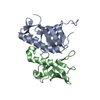
Crystal Structure of Full-length Human RPA 14/32 Heterodimer
Components
Replication protein A 14 kDa subunit
Replication protein A 32 kDa subunit
Keywords
REPLICATION / RPA14/32 / ssDNA binding protein / OB-fold
Function / homology
Function and homology information
protein localization to chromosome / DNA replication factor A complex / Removal of the Flap Intermediate / G-rich strand telomeric DNA binding / Mismatch repair (MMR) directed by MSH2:MSH3 (MutSbeta) / Mismatch repair (MMR) directed by MSH2:MSH6 (MutSalpha) / regulation of DNA damage checkpoint / Removal of the Flap Intermediate from the C-strand / regulation of double-strand break repair via homologous recombination / HDR through Single Strand Annealing (SSA) ...protein localization to chromosome / DNA replication factor A complex / Removal of the Flap Intermediate / G-rich strand telomeric DNA binding / Mismatch repair (MMR) directed by MSH2:MSH3 (MutSbeta) / Mismatch repair (MMR) directed by MSH2:MSH6 (MutSalpha) / regulation of DNA damage checkpoint / Removal of the Flap Intermediate from the C-strand / regulation of double-strand break repair via homologous recombination / HDR through Single Strand Annealing (SSA) / telomeric repeat DNA binding / Impaired BRCA2 binding to RAD51 / PCNA-Dependent Long Patch Base Excision Repair / Activation of the pre-replicative complex / Regulation of HSF1-mediated heat shock response / Presynaptic phase of homologous DNA pairing and strand exchange / HSF1 activation / Activation of ATR in response to replication stress / mismatch repair / mitotic G1 DNA damage checkpoint signaling / regulation of mitotic cell cycle / telomere maintenance / Translesion synthesis by REV1 / Translesion synthesis by POLK / Translesion synthesis by POLI / Gap-filling DNA repair synthesis and ligation in GG-NER / nucleotide-excision repair / Fanconi Anemia Pathway / Termination of translesion DNA synthesis / Translesion Synthesis by POLH / base-excision repair / PML body / Recognition of DNA damage by PCNA-containing replication complex / G2/M DNA damage checkpoint / double-strand break repair via homologous recombination / Meiotic recombination / HDR through Homologous Recombination (HRR) / Dual Incision in GG-NER / Formation of Incision Complex in GG-NER / Dual incision in TC-NER / Gap-filling DNA repair synthesis and ligation in TC-NER / regulation of cell population proliferation / single-stranded DNA binding / site of double-strand break / Processing of DNA double-strand break ends / protein phosphatase binding / Regulation of TP53 Activity through Phosphorylation / damaged DNA binding / chromosome, telomeric region / DNA replication / nuclear body / DNA repair / ubiquitin protein ligase binding / chromatin / enzyme binding / nucleoplasm / nucleus Similarity search - Function
Replication factor A protein 2 / Replication protein A, C-terminal / Replication protein A C terminal / Replication factor A protein 3 / Replication factor A protein 3 / Replication factor A protein-like / Nucleic acid-binding proteins / OB fold (Dihydrolipoamide Acetyltransferase, E2P) / Winged helix DNA-binding domain superfamily / Winged helix-like DNA-binding domain superfamily ...Replication factor A protein 2 / Replication protein A, C-terminal / Replication protein A C terminal / Replication factor A protein 3 / Replication factor A protein 3 / Replication factor A protein-like / Nucleic acid-binding proteins / OB fold (Dihydrolipoamide Acetyltransferase, E2P) / Winged helix DNA-binding domain superfamily / Winged helix-like DNA-binding domain superfamily / Nucleic acid-binding, OB-fold / Beta Barrel / Mainly Beta Similarity search - Domain/homology
A: Replication protein A 32 kDa subunit B: Replication protein A 14 kDa subunit C: Replication protein A 32 kDa subunit D: Replication protein A 14 kDa subunit
Resolution: 2.4→2.58 Å / Redundancy: 5.9 % / Mean I/σ(I) obs: 3.8 / Num. unique all: 3730 / Rsym value: 0.58 / % possible all: 97.3
-
Processing
Software
Name
Version
Classification
REFMAC
5.2.0005
refinement
HKL-2000
datacollection
HKL-2000
datareduction
HKL-2000
datascaling
SHELXS
phasing
Refinement
Method to determine structure: MAD / Resolution: 2.5→25 Å / Cor.coef. Fo:Fc: 0.924 / Cor.coef. Fo:Fc free: 0.884 / SU B: 10.578 / SU ML: 0.237 / Cross valid method: THROUGHOUT / σ(F): 3 / σ(I): 3 / ESU R: 0.551 / ESU R Free: 0.318 / Stereochemistry target values: MAXIMUM LIKELIHOOD / Details: High R value due to the disordered domain
Rfactor
Num. reflection
% reflection
Selection details
Rfree
0.27981
1049
5.1 %
RANDOM
Rwork
0.22825
-
-
-
obs
0.23091
19411
96.03 %
-
all
-
20156
-
-
Solvent computation
Ion probe radii: 0.8 Å / Shrinkage radii: 0.8 Å / VDW probe radii: 1.2 Å / Solvent model: MASK
Displacement parameters
Biso mean: 37.724 Å2
Baniso -1
Baniso -2
Baniso -3
1-
1.69 Å2
0.84 Å2
0 Å2
2-
-
1.69 Å2
0 Å2
3-
-
-
-2.53 Å2
Refinement step
Cycle: LAST / Resolution: 2.5→25 Å
Protein
Nucleic acid
Ligand
Solvent
Total
Num. atoms
3785
0
0
0
3785
Refine LS restraints
Refine-ID
Type
Dev ideal
Dev ideal target
Number
X-RAY DIFFRACTION
r_bond_refined_d
0.012
0.022
3850
X-RAY DIFFRACTION
r_angle_refined_deg
1.505
1.965
5220
X-RAY DIFFRACTION
r_dihedral_angle_1_deg
7.534
5
477
X-RAY DIFFRACTION
r_dihedral_angle_2_deg
36.374
24.847
163
X-RAY DIFFRACTION
r_dihedral_angle_3_deg
18.503
15
687
X-RAY DIFFRACTION
r_dihedral_angle_4_deg
16.583
15
17
X-RAY DIFFRACTION
r_chiral_restr
0.113
0.2
617
X-RAY DIFFRACTION
r_gen_planes_refined
0.004
0.02
2831
X-RAY DIFFRACTION
r_nbd_refined
0.23
0.2
1580
X-RAY DIFFRACTION
r_nbtor_refined
0.31
0.2
2595
X-RAY DIFFRACTION
r_xyhbond_nbd_refined
0.146
0.2
111
X-RAY DIFFRACTION
r_symmetry_vdw_refined
0.192
0.2
47
X-RAY DIFFRACTION
r_symmetry_hbond_refined
0.197
0.2
6
X-RAY DIFFRACTION
r_mcbond_it
0.71
1.5
2472
X-RAY DIFFRACTION
r_mcangle_it
1.238
2
3938
X-RAY DIFFRACTION
r_scbond_it
1.447
3
1526
X-RAY DIFFRACTION
r_scangle_it
2.291
4.5
1282
LS refinement shell
Resolution: 2.5→2.564 Å / Total num. of bins used: 20
Rfactor
Num. reflection
% reflection
Rfree
0.33
76
-
Rwork
0.31
1428
-
obs
-
1525
97.22 %
+
About Yorodumi
-
News
-
Feb 9, 2022. New format data for meta-information of EMDB entries
New format data for meta-information of EMDB entries
Version 3 of the EMDB header file is now the official format.
The previous official version 1.9 will be removed from the archive.
In the structure databanks used in Yorodumi, some data are registered as the other names, "COVID-19 virus" and "2019-nCoV". Here are the details of the virus and the list of structure data.
Jan 31, 2019. EMDB accession codes are about to change! (news from PDBe EMDB page)
EMDB accession codes are about to change! (news from PDBe EMDB page)
The allocation of 4 digits for EMDB accession codes will soon come to an end. Whilst these codes will remain in use, new EMDB accession codes will include an additional digit and will expand incrementally as the available range of codes is exhausted. The current 4-digit format prefixed with “EMD-” (i.e. EMD-XXXX) will advance to a 5-digit format (i.e. EMD-XXXXX), and so on. It is currently estimated that the 4-digit codes will be depleted around Spring 2019, at which point the 5-digit format will come into force.
The EM Navigator/Yorodumi systems omit the EMD- prefix.
Related info.:Q: What is EMD? / ID/Accession-code notation in Yorodumi/EM Navigator
Yorodumi is a browser for structure data from EMDB, PDB, SASBDB, etc.
This page is also the successor to EM Navigator detail page, and also detail information page/front-end page for Omokage search.
The word "yorodu" (or yorozu) is an old Japanese word meaning "ten thousand". "mi" (miru) is to see.
Related info.:EMDB / PDB / SASBDB / Comparison of 3 databanks / Yorodumi Search / Aug 31, 2016. New EM Navigator & Yorodumi / Yorodumi Papers / Jmol/JSmol / Function and homology information / Changes in new EM Navigator and Yorodumi
 Movie
Movie Controller


 Open data
 Basic information
 Components
 Keywords
 Function and homology information
 Homo sapiens (human)
 X-RAY DIFFRACTION /
  Authors
 Citation
 Structure visualization
 Downloads & links
 Other downloads


 PDBj
PDBj


 Assembly
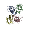


 Sample preparation
 / Beamline: 17-ID / Wavelength: 0.9794728, 0.979609, 0.999879
 Processing