 Movie
Movie Controller


+ Open data

- Basic information
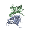
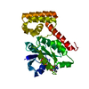
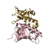
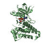
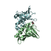
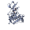
| Entry | Database: PDB / ID: 2z6k | ||||||
|---|---|---|---|---|---|---|---|
| Title | Crystal structure of full-length human RPA14/32 heterodimer | ||||||
 Components Components |
| ||||||
 Keywords Keywords | REPLICATION / full-length RPA14/32 / ssDNA binding protein / OB-fold / Acetylation / Alternative splicing / DNA replication / Nucleus / Phosphorylation / Polymorphism | ||||||
| Function / homology |  Function and homology information Function and homology informationprotein localization to chromosome / DNA replication factor A complex / Removal of the Flap Intermediate / G-rich strand telomeric DNA binding / Mismatch repair (MMR) directed by MSH2:MSH3 (MutSbeta) / Mismatch repair (MMR) directed by MSH2:MSH6 (MutSalpha) / regulation of DNA damage checkpoint / Removal of the Flap Intermediate from the C-strand / regulation of double-strand break repair via homologous recombination / HDR through Single Strand Annealing (SSA) ...protein localization to chromosome / DNA replication factor A complex / Removal of the Flap Intermediate / G-rich strand telomeric DNA binding / Mismatch repair (MMR) directed by MSH2:MSH3 (MutSbeta) / Mismatch repair (MMR) directed by MSH2:MSH6 (MutSalpha) / regulation of DNA damage checkpoint / Removal of the Flap Intermediate from the C-strand / regulation of double-strand break repair via homologous recombination / HDR through Single Strand Annealing (SSA) / telomeric repeat DNA binding / Impaired BRCA2 binding to RAD51 / PCNA-Dependent Long Patch Base Excision Repair / Activation of the pre-replicative complex / Regulation of HSF1-mediated heat shock response / Presynaptic phase of homologous DNA pairing and strand exchange / HSF1 activation / Activation of ATR in response to replication stress / mismatch repair / mitotic G1 DNA damage checkpoint signaling / telomere maintenance / Translesion synthesis by REV1 / Translesion synthesis by POLK / Translesion synthesis by POLI / Gap-filling DNA repair synthesis and ligation in GG-NER / nucleotide-excision repair / Fanconi Anemia Pathway / Termination of translesion DNA synthesis / Translesion Synthesis by POLH / base-excision repair / PML body / Recognition of DNA damage by PCNA-containing replication complex / double-strand break repair via homologous recombination / G2/M DNA damage checkpoint / Meiotic recombination / HDR through Homologous Recombination (HRR) / Dual Incision in GG-NER / Formation of Incision Complex in GG-NER / Dual incision in TC-NER / Gap-filling DNA repair synthesis and ligation in TC-NER / single-stranded DNA binding / site of double-strand break / Processing of DNA double-strand break ends / protein phosphatase binding / Regulation of TP53 Activity through Phosphorylation / damaged DNA binding / nuclear body / chromosome, telomeric region / DNA replication / DNA repair / ubiquitin protein ligase binding / chromatin / enzyme binding / nucleoplasm / nucleus Similarity search - Function | ||||||
| Biological species |  Homo sapiens (human) Homo sapiens (human) | ||||||
| Method |  X-RAY DIFFRACTION / MOLECULAR REPLACEMENT / Resolution: 3 Å X-RAY DIFFRACTION / MOLECULAR REPLACEMENT / Resolution: 3 Å | ||||||
 Authors Authors | Deng, X. / Habel, J.E. / Kabaleeswaran, V. / Borgstahl, G.E. | ||||||
 Citation Citation | Journal: J.Mol.Biol. / Year: 2007 Title: Structure of the Full-length Human RPA14/32 Complex Gives Insights into the Mechanism of DNA Binding and Complex Formation Authors: Deng, X. / Habel, J.E. / Kabaleeswaran, V. / Snell, E.H. / Wold, M.S. / Borgstahl, G.E. | ||||||
| History |
|
- Structure visualization
| Structure viewer | Molecule: MolmilJmol/JSmol |
|---|
- Downloads & links
-Download
| PDBx/mmCIF format | 2z6k.cif.gz | 110.8 KB | Display | PDBx/mmCIF format |
|---|---|---|---|---|
| PDB format | pdb2z6k.ent.gz | 83.8 KB | Display | PDB format |
| PDBx/mmJSON format | 2z6k.json.gz | Tree view | PDBx/mmJSON format | |
| Others |  Other downloads Other downloads |
-Validation report
| Arichive directory | https://data.pdbj.org/pub/pdb/validation_reports/z6/2z6kftp://data.pdbj.org/pub/pdb/validation_reports/z6/2z6k | HTTPS FTP |
|---|
-Related structure data
| Related structure data |  2pi2C  2pqaC 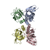 1quqS C: citing same article ( S: Starting model for refinement |
|---|---|
| Similar structure data |


-Links
 PDBj
PDBj


- Assembly
| Deposited unit | 
| ||||||||
|---|---|---|---|---|---|---|---|---|---|
| 1 | 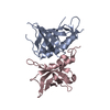
| ||||||||
| 2 | 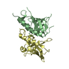
| ||||||||
| Unit cell |
|
-Components
| #1: Protein | Mass: 29276.795 Da / Num. of mol.: 2 / Fragment: residues 42-175 Source method: isolated from a genetically manipulated source Source: (gene. exp.) Homo sapiens (human) / Gene: RPA2, REPA2, RPA32 / Plasmid: pET16b / Production host:  #2: Protein | Mass: 16117.456 Da / Num. of mol.: 2 Source method: isolated from a genetically manipulated source Source: (gene. exp.) Homo sapiens (human) / Gene: RPA3, REPA3, RPA14 / Plasmid: pET16b / Production host: |
|---|
-Experimental details
-Experiment
| Experiment | Method: X-RAY DIFFRACTION / Number of used crystals: 1 |
|---|
- Sample preparation
| Crystal | Density Matthews: 3.286145 Å3/Da / Density % sol: 62.570122 % |
|---|---|
| Crystal grow | Temperature: 293 K / Method: evaporation / pH: 5.9 Details: 0.95M KNa tartrate, 0.1M MES, 10mM DTT, pH 5.9, EVAPORATION, temperature 293K |
-Data collection
| Diffraction | Mean temperature: 100 K |
|---|---|
| Diffraction source | Source: ROTATING ANODE / Type: RIGAKU FR-E+ SUPERBRIGHT / Wavelength: 1.5418 |
| Detector | Type: RIGAKU RAXIS IV++ / Detector: IMAGE PLATE / Date: Feb 24, 2004 |
| Radiation | Protocol: SINGLE WAVELENGTH / Monochromatic (M) / Laue (L): M / Scattering type: x-ray |
| Radiation wavelength | Wavelength: 1.5418 Å / Relative weight: 1 |
| Reflection | Resolution: 2.9→50 Å / Num. all: 26620 / Num. obs: 25484 / % possible obs: 95.8 % / Observed criterion σ(F): 1 / Observed criterion σ(I): 1 / Redundancy: 3.1 % / Biso Wilson estimate: 90 Å2 / Rsym value: 0.07 / Net I/σ(I): 16 |
| Reflection shell | Resolution: 2.98→3.1 Å / Redundancy: 3.2 % / Mean I/σ(I) obs: 1.6 / Num. unique all: 2647 / Rsym value: 0.7 / % possible all: 99 |
- Processing
| Software |
| ||||||||||||||||||||||||||||||||||||||||||||||||||||||||||||||||||||||||||||||||||||||||||||||||||||||||||||||||||||||||||||||||||||||||||||||||||||||||||||||||||||||||||
|---|---|---|---|---|---|---|---|---|---|---|---|---|---|---|---|---|---|---|---|---|---|---|---|---|---|---|---|---|---|---|---|---|---|---|---|---|---|---|---|---|---|---|---|---|---|---|---|---|---|---|---|---|---|---|---|---|---|---|---|---|---|---|---|---|---|---|---|---|---|---|---|---|---|---|---|---|---|---|---|---|---|---|---|---|---|---|---|---|---|---|---|---|---|---|---|---|---|---|---|---|---|---|---|---|---|---|---|---|---|---|---|---|---|---|---|---|---|---|---|---|---|---|---|---|---|---|---|---|---|---|---|---|---|---|---|---|---|---|---|---|---|---|---|---|---|---|---|---|---|---|---|---|---|---|---|---|---|---|---|---|---|---|---|---|---|---|---|---|---|---|---|
| Refinement | Method to determine structure: MOLECULAR REPLACEMENT Starting model: PDB ENTRY 1QUQ Resolution: 3→50 Å / Cor.coef. Fo:Fc: 0.938 / Cor.coef. Fo:Fc free: 0.914 / SU B: 15.237 / SU ML: 0.273 / Cross valid method: THROUGHOUT / σ(F): 2 / σ(I): 2 / ESU R: 0.511 / ESU R Free: 0.339 / Stereochemistry target values: MAXIMUM LIKELIHOOD
| ||||||||||||||||||||||||||||||||||||||||||||||||||||||||||||||||||||||||||||||||||||||||||||||||||||||||||||||||||||||||||||||||||||||||||||||||||||||||||||||||||||||||||
| Solvent computation | Ion probe radii: 0.8 Å / Shrinkage radii: 0.8 Å / VDW probe radii: 1.2 Å / Solvent model: MASK | ||||||||||||||||||||||||||||||||||||||||||||||||||||||||||||||||||||||||||||||||||||||||||||||||||||||||||||||||||||||||||||||||||||||||||||||||||||||||||||||||||||||||||
| Displacement parameters | Biso mean: 89.456 Å2
| ||||||||||||||||||||||||||||||||||||||||||||||||||||||||||||||||||||||||||||||||||||||||||||||||||||||||||||||||||||||||||||||||||||||||||||||||||||||||||||||||||||||||||
| Refinement step | Cycle: LAST / Resolution: 3→50 Å
| ||||||||||||||||||||||||||||||||||||||||||||||||||||||||||||||||||||||||||||||||||||||||||||||||||||||||||||||||||||||||||||||||||||||||||||||||||||||||||||||||||||||||||
| Refine LS restraints |
| ||||||||||||||||||||||||||||||||||||||||||||||||||||||||||||||||||||||||||||||||||||||||||||||||||||||||||||||||||||||||||||||||||||||||||||||||||||||||||||||||||||||||||
| LS refinement shell | Resolution: 3→3.078 Å / Total num. of bins used: 20
|
